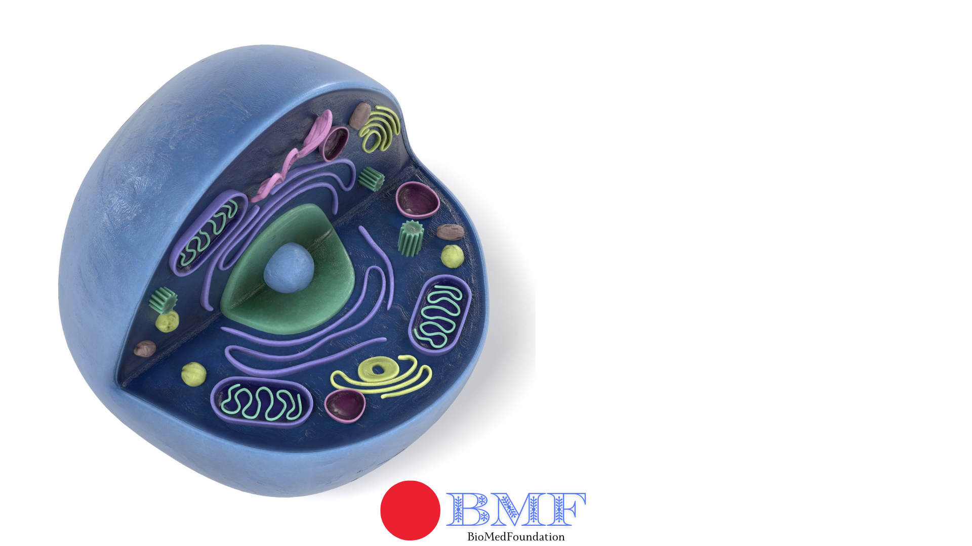
4.01 Overview: Cell Membrane
Role and Presence:
- Acts as a boundary and barrier.
- Present in both eukaryotic and prokaryotic cells
- Defines the cell’s internal and external environments.
- Forms compartments within cells by surrounding organelles (e.g., nucleus, mitochondria, and rough endoplasmic reticulum).
Key Functions:
- Transport Mechanisms:
- Materials cross membranes via:
- diffusion
- osmosis
- active transport
- bulk transport
- Materials cross membranes via:
- Selective Permeability:
- Controls entry and exit of substances.
- Communication:
- Acts as an interface for:
- cell signalling
- allowing chemical messaging (e.g., hormones, growth factors)
- identity recognition.
- Acts as an interface for:
Membrane Permeability
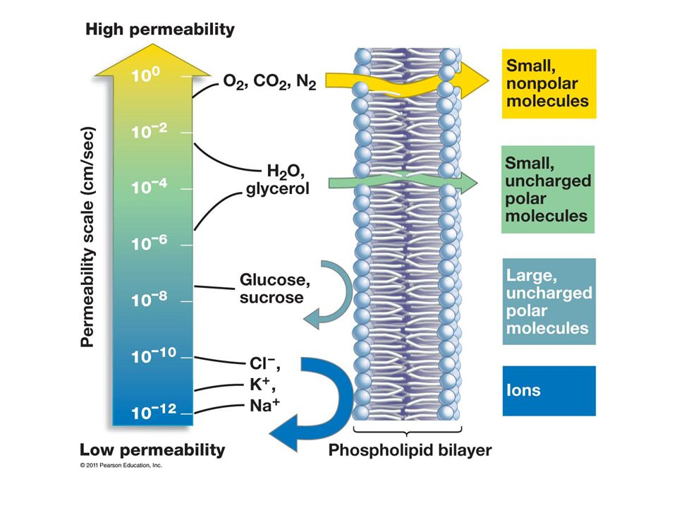
1. Selective Permeability
- The cell membrane is selectively permeable, meaning it only allows certain substances to cross freely while restricting others.
- Unassisted Passage:
- Small
- Nonpolar
- Uncharged
- Lipid-soluble molecules
- Example: oxygen (O₂) and carbon dioxide (CO₂), can diffuse through the phospholipid bilayer without assistance.
- Water molecules pass slowly due to their polarity but can move more freely through special channels called aquaporins.
- Water-Soluble Molecules:
- Large or polar (water-soluble) molecules face difficulty passing through the membrane due to the hydrophobic (water-repelling) core of the bilayer, which acts as a barrier.
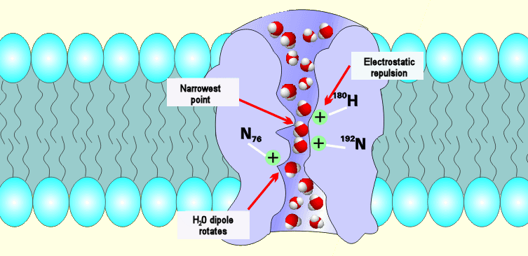
Figure: Aquaporin
2. Barrier Properties
- The lipid bilayer structure primarily blocks polar molecules and ions from crossing freely, maintaining the internal environment of the cell.
- Hydrophobic Core:
- The hydrophobic tails of phospholipids create a nonpolar barrier, preventing polar and charged particles, such as ions and sugars, from easily passing through.
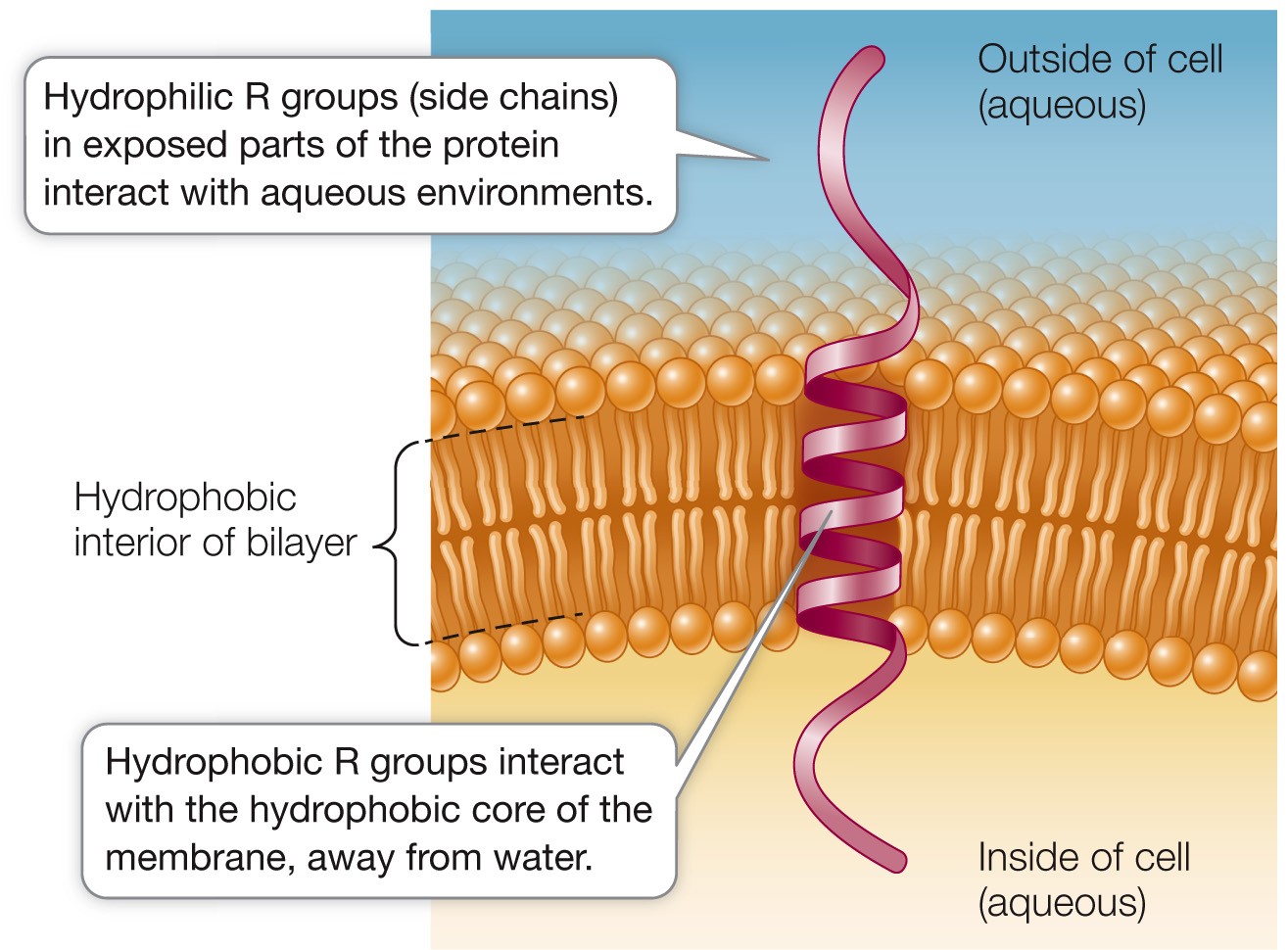
- Transport Proteins:
- To facilitate the movement of these substances, the membrane utilizes channel and carrier proteins that selectively transport ions, glucose, amino acids, and other necessary molecules across the membrane.
Fluid Mosaic Model

Diagram above: An example of Mosaic art
The Fluid Mosaic Model:
- It is a widely accepted scientific model that describes the structure of cell membranes.
- The model illustrates how cell membranes are not rigid, but rather flexible and dynamic.
Protein Distribution:
- Proteins are scattered within the phospholipid bilayer, forming a mosaic-like pattern.
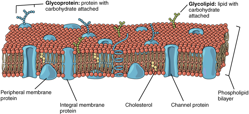
“Fluid” Component:
- Molecular Movement: Phospholipids and proteins move laterally, maintaining a fluid state with consistency similar to olive oil.
“Mosaic” Component:
- Protein Distribution: Proteins are scattered within the phospholipid bilayer, forming a mosaic-like pattern.
Compartmentalization and Division of Labour
Division of Labour
Importance of Compartmentalization
- Specialization: Allows organelles to maintain environments suited to their functions (e.g., lysosomes maintain an acidic pH for enzyme activity).
- Efficiency: Isolates biochemical processes, preventing interference and optimizing reaction conditions.
Practise Questions
Question 1
Describe the structure and role of the cell membrane in both eukaryotic and prokaryotic cells. (6 marks)
Mark Scheme:
- The cell membrane is a phospholipid bilayer present in both eukaryotic and prokaryotic cells. (1 mark)
- It acts as a boundary that separates the internal environment of the cell from the external environment. (1 mark)
- The membrane provides compartmentalization by surrounding organelles in eukaryotic cells (e.g., nucleus, mitochondria). (1 mark)
- It is selectively permeable, controlling the entry and exit of substances. (1 mark)
- It facilitates cell communication through signalling and recognition (e.g., hormone receptors). (1 mark)
- It allows materials to cross via diffusion, osmosis, active transport, and bulk transport mechanisms. (1 mark)
Question 2
Explain the concept of selective permeability and its importance to the cell membrane. (5 marks)
Mark Scheme:
- Selective permeability means the cell membrane allows certain substances to pass while restricting others. (1 mark)
- Small, nonpolar, uncharged, lipid-soluble molecules (e.g., O₂, CO₂) can diffuse freely through the phospholipid bilayer. (1 mark)
- Polar or large water-soluble molecules require transport proteins (e.g., glucose and ions). (1 mark)
- The hydrophobic core of the lipid bilayer prevents polar and charged substances from crossing freely. (1 mark)
- This property helps maintain the internal environment of the cell by regulating molecular exchange. (1 mark)
Question 3
How does the fluid mosaic model describe the structure of cell membranes? (6 marks)
Mark Scheme:
- The fluid mosaic model describes the cell membrane as a flexible and dynamic structure. (1 mark)
- It consists of a phospholipid bilayer with embedded proteins, forming a mosaic-like pattern. (1 mark)
- The “fluid” component refers to the lateral movement of phospholipids and proteins within the bilayer. (1 mark)
- This fluidity allows the membrane to adapt to changes and maintain its integrity. (1 mark)
- The “mosaic” component refers to the distribution of proteins scattered within the bilayer. (1 mark)
- Examples of proteins include channel proteins, carrier proteins, and receptors involved in transport and communication. (1 mark)
Question 4
Describe the role of transport proteins in the cell membrane. (5 marks)
Mark Scheme:
- Transport proteins facilitate the movement of substances that cannot pass freely through the lipid bilayer. (1 mark)
- Channel proteins form hydrophilic pores, allowing ions and small polar molecules to cross the membrane. (1 mark)
- Carrier proteins bind to specific molecules (e.g., glucose, amino acids) and change shape to transport them across the membrane. (1 mark)
- These proteins enable the selective transport of essential substances while maintaining the barrier properties of the membrane. (1 mark)
- They play a key role in active transport and facilitated diffusion. (1 mark)
Question 5
How does the hydrophobic core of the lipid bilayer affect membrane permeability? (5 marks)
Mark Scheme:
- The hydrophobic core is formed by the fatty acid tails of phospholipids, which are nonpolar and water-repelling. (1 mark)
- It creates a barrier that prevents the free passage of polar molecules and ions (e.g., Na⁺, Cl⁻). (1 mark)
- Small, nonpolar molecules like O₂ and CO₂ can diffuse through the core without assistance. (1 mark)
- Larger or polar molecules require specific transport proteins to cross the membrane. (1 mark)
- This selective permeability ensures the cell maintains its internal environment. (1 mark)
Question 6
Explain the significance of aquaporins in the cell membrane. (4 marks)
Mark Scheme:
- Aquaporins are specialized channel proteins that facilitate the movement of water across the cell membrane. (1 mark)
- They allow water molecules to pass through the membrane more efficiently than by simple diffusion. (1 mark)
- This is especially important for cells involved in osmosis or rapid water transport (e.g., kidney cells). (1 mark)
- Aquaporins help maintain water balance within the cell, supporting proper cellular function. (1 mark)
Question 7
What is the importance of membrane fluidity, and how is it maintained? (6 marks)
Mark Scheme:
- Membrane fluidity allows the membrane to remain flexible and adapt to changes in the environment. (1 mark)
- It ensures proper functioning of proteins involved in transport, signalling, and recognition. (1 mark)
- Lateral movement of phospholipids within the bilayer maintains the fluid state. (1 mark)
- The presence of cholesterol helps regulate fluidity by preventing the membrane from becoming too rigid or too fluid. (1 mark)
- Unsaturated fatty acid tails in phospholipids increase fluidity by preventing tight packing. (1 mark)
- Fluidity is crucial for processes like endocytosis, exocytosis, and membrane repair. (1 mark)
Question 8
Explain how the cell membrane acts as an interface for cell signalling. (5 marks)
Mark Scheme:
- The cell membrane contains receptor proteins that bind to signalling molecules like hormones and growth factors. (1 mark)
- These receptors detect specific signals and initiate intracellular responses. (1 mark)
- Example: Insulin binds to its receptor, triggering glucose uptake in cells. (1 mark)
- The membrane also facilitates identity recognition, such as immune cells identifying pathogens. (1 mark)
- This communication is essential for maintaining homeostasis and coordinating cellular activities. (1 mark)
Question 9
Why is the fluid mosaic model considered dynamic, and how does this benefit the cell? (5 marks)
Mark Scheme:
- The model is dynamic because phospholipids and proteins can move laterally within the bilayer. (1 mark)
- This movement allows the membrane to adapt to environmental changes, such as temperature fluctuations. (1 mark)
- The flexibility supports membrane processes like vesicle formation, endocytosis, and exocytosis. (1 mark)
- Protein movement within the bilayer ensures proper function in transport and signalling. (1 mark)
- The dynamic nature allows cells to maintain structural integrity while remaining responsive to their environment. (1 mark)
Question 10
Discuss the significance of the hydrophilic and hydrophobic regions of the phospholipid bilayer in maintaining membrane structure. (6 marks)
Mark Scheme:
- The hydrophilic heads of phospholipids face outward, interacting with water inside and outside the cell. (1 mark)
- The hydrophobic tails face inward, forming a nonpolar core that prevents water-soluble substances from crossing freely. (1 mark)
- This arrangement creates a stable bilayer structure. (1 mark)
- It ensures selective permeability, allowing the membrane to control molecular exchange. (1 mark)
- The hydrophilic and hydrophobic regions maintain the barrier properties while supporting protein integration. (1 mark)
- This dual nature enables the membrane to be both flexible and functional. (1 mark)
Exam Style Questions
Quiz 1
1. What does it mean for the cell membrane to be selectively permeable?
2. Why do large or polar molecules face difficulty passing through the cell membrane?
3. Which type of molecules can diffuse unassisted across the phospholipid bilayer?
4. How do water molecules cross the cell membrane more easily, despite being polar?
5. Which of the following molecules would most likely require a transport protein to cross the cell membrane?
Correct Answers: 0%
Exam Style Questions
Test 1
1. Which of the following best describes the role of a cell membrane?
2. What characteristic of the cell membrane enables selective permeability?
3. Which transport process requires energy in the form of ATP?
4. Which structure is found in both eukaryotic and prokaryotic cells?
5. Which process involves the movement of water across a selectively permeable membrane?
6. Which cellular process is essential for cell signaling at the cell membrane?
7. Which type of molecule is primarily responsible for forming the cell membrane’s structure?
8. How does bulk transport across the cell membrane differ from diffusion?
9. In cell signaling, which molecules typically act as chemical messengers that bind to membrane receptors?
10. Which of the following best describes the fluid-mosaic model of the cell membrane?
Correct Answers: 0%