4.02 Phospholipid Bilayer
1. Structure of Phospholipids
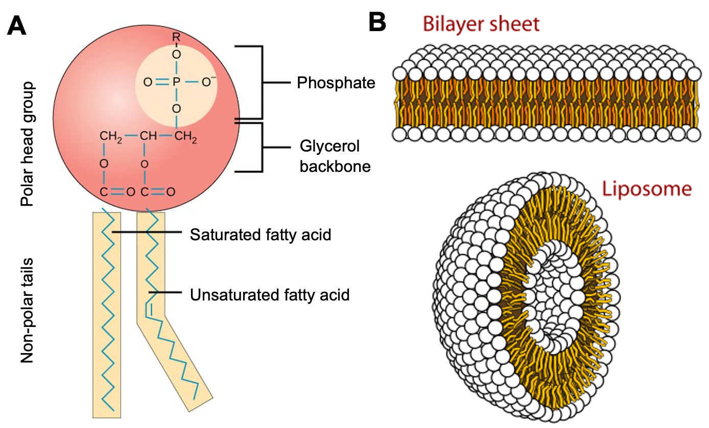
- Basic Components:
- Glycerol Backbone:
- A three-carbon molecule serving as the core.
- Two Fatty Acid Tails:
- Long hydrocarbon chains attached to the glycerol.
- These are hydrophobic (water-repelling).
- Phosphate Group Head:
- Attached to the third carbon of glycerol.
- Often linked to additional molecules (e.g., choline).
- This head is hydrophilic (water-attracting).
- Glycerol Backbone:
- Amphipathic Nature:
- Phospholipids have both hydrophilic and hydrophobic regions, giving them a unique arrangement in water environments.
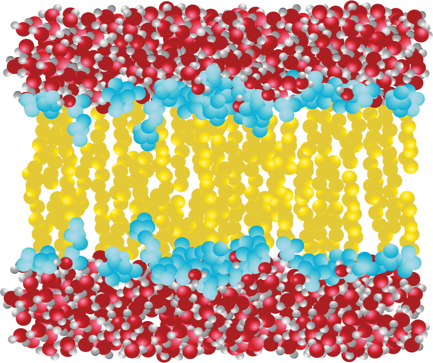
- Arrangement in Bilayer:
- Phospholipids form a bilayer in cell membranes, with hydrophobic tails facing inward and hydrophilic heads facing outward towards the water-based environment, both inside and outside the cell.

2. Properties of Phospholipids
- Hydrophobic Tails: Nonpolar, repel water, and orient away from water molecules.
- Hydrophilic Head: Polar, attracts water, and interacts with water molecules.
- Self-Assembly:
- In aqueous solutions, phospholipids spontaneously arrange into structures like bilayers, with heads facing outwards and tails facing inwards, away from water.
3. Role in Cell Membranes
- Phospholipid Bilayer:
- Forms the basic structure of the cell membrane, creating a selectively permeable barrier.
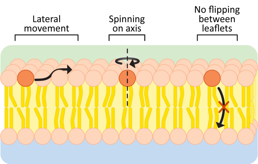
- Fluidity and Flexibility:
- Allows for membrane fluidity; phospholipids move laterally within the layer, crucial for membrane functions like cell signalling, endocytosis, and exocytosis.
- Barrier Function:
- Prevents most water-soluble molecules (e.g., sugars, amino acids, ions) from freely passing across the membrane.
- Maintains cellular integrity by preventing the leakage of essential molecules and blocking the entry of unwanted substances.
4. Signalling Functions of Phospholipids
- Signalling Molecules:
- Can be hydrolysed to release small, water-soluble molecules that act as signalling molecules within cells.
- These molecules bind to specific receptors in the cytoplasm to trigger cellular responses.
- Activation of Enzymes:
- Certain phospholipids can move within the bilayer to interact with and activate other molecules, such as enzymes, that play roles in cellular signalling pathways.
5. Other Functions of Phospholipids
- Formation of Vesicles:
- Phospholipids enable the formation of vesicles, which transport materials within cells.
- Cellular Communication:
- Some phospholipids act as intracellular messengers, such as phosphatidylinositol, which is involved in signal transduction pathways.
Structure of the Phospholipid Bilayer

1. Structure of the Phospholipid Bilayer
- Cholesterol:
- Interspersed among phospholipids
- Stabilises the membrane by limiting lipid movement and reducing fluidity.
- Thickness:
- Less than 10 nm
- Visible only under an electron microscope.
2. Composition and Arrangement:
- Phospholipids:
- Form a bilayer
- Hydrophilic (water-attracting) heads facing outward
- Hydrophobic (water-repelling) tails facing inward.
- Self-Assembly in Water:
- Monolayer:
- Phospholipids arrange with heads in water, tails sticking out.
- Micelle:
- Spherical formation with tails inward and heads outward.
- Number 2 in the figure below.
- Bilayer:
- Hydrophobic tails face each other
- Creates a stable, two-layer barrier.
- Number 1 in the figure below.
- Monolayer:
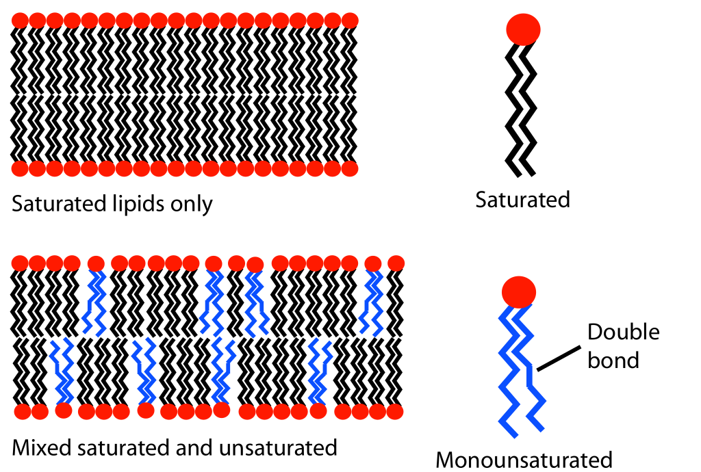
- Fluidity Control:
- Saturated Fatty Acids:
- Have straight tails that pack closely, reducing fluidity.
- Unsaturated Fatty Acids:
- Kinked tails prevent close packing, increasing fluidity.
- Saturated Fatty Acids:


3. Barrier Function:
- Prevents most water-soluble molecules (e.g., sugars, amino acids, ions) from passing across the membrane.
- Maintains cellular integrity by preventing leakage of essential molecules and entry of unwanted substances.
4. Signalling:
- Can move within the bilayer.
- Distributes around cell receptors.
- External signals can bind to theses receptors and activate other molecules like enzymes inside the cell.
Practise Questions
Question 1
Describe the structure of a phospholipid and explain its amphipathic nature. (6 marks)
Mark Scheme:
- A phospholipid consists of a glycerol backbone attached to two fatty acid tails and a phosphate group. (1 mark)
- The fatty acid tails are long hydrocarbon chains, hydrophobic and water-repelling. (1 mark)
- The phosphate group forms a hydrophilic, water-attracting head. (1 mark)
- Phospholipids are amphipathic, meaning they have both hydrophilic (polar) and hydrophobic (nonpolar) regions. (1 mark)
- In an aqueous environment, phospholipids arrange themselves with tails inward and heads outward, forming bilayers or micelles. (1 mark)
- This unique arrangement is essential for forming biological membranes. (1 mark)
Question 2
Explain how the amphipathic nature of phospholipids contributes to their role in cell membranes. (5 marks)
Mark Scheme:
- The hydrophilic heads face the aqueous environments inside and outside the cell. (1 mark)
- The hydrophobic tails face inward, away from water, creating a nonpolar core. (1 mark)
- This arrangement forms a bilayer, which is selectively permeable. (1 mark)
- The bilayer prevents the free passage of water-soluble molecules (e.g., ions, sugars), maintaining cellular integrity. (1 mark)
- The amphipathic nature allows for membrane fluidity and flexibility, supporting processes like endocytosis, exocytosis, and signalling. (1 mark)
Question 3
Outline the role of phospholipids in the barrier and signalling functions of the cell membrane. (6 marks)
Mark Scheme:
- Barrier Function: Phospholipids form a bilayer that prevents most water-soluble molecules (e.g., ions, amino acids) from crossing freely. (1 mark)
- The hydrophobic core ensures that only small, nonpolar molecules (e.g., O₂, CO₂) can diffuse freely. (1 mark)
- Signalling Function: Phospholipids can be hydrolysed to release small signalling molecules that trigger intracellular responses. (1 mark)
- They can move laterally within the bilayer to interact with and activate other molecules, such as enzymes. (1 mark)
- Example: Phosphatidylinositol plays a role in signal transduction pathways. (1 mark)
- These properties allow cells to respond to external stimuli efficiently. (1 mark)
Question 4
Describe the structure and significance of the phospholipid bilayer. (6 marks)
Mark Scheme:
- The bilayer consists of two layers of phospholipids with hydrophobic tails facing inward and hydrophilic heads facing outward. (1 mark)
- The arrangement creates a stable barrier that separates the cell’s internal and external environments. (1 mark)
- The bilayer is fluid, with phospholipids and proteins able to move laterally within the membrane. (1 mark)
- Cholesterol interspersed among phospholipids stabilizes the membrane by controlling fluidity. (1 mark)
- The bilayer’s barrier properties prevent the leakage of essential molecules and block unwanted substances. (1 mark)
- The bilayer supports membrane functions such as transport, signalling, and compartmentalization. (1 mark)
Question 5
How do saturated and unsaturated fatty acids affect membrane fluidity? (4 marks)
Mark Scheme:
- Saturated fatty acids have straight tails that pack closely together, reducing membrane fluidity. (1 mark)
- Unsaturated fatty acids have kinked tails due to double bonds, preventing tight packing and increasing fluidity. (1 mark)
- Membrane fluidity is crucial for processes like endocytosis, exocytosis, and protein function. (1 mark)
- The balance between saturated and unsaturated fatty acids ensures optimal membrane flexibility and stability. (1 mark)
Question 6
Explain how phospholipids contribute to the formation of vesicles and their role in cellular transport. (5 marks)
Mark Scheme:
- Phospholipids enable the spontaneous formation of vesicles by enclosing materials in a bilayer. (1 mark)
- The hydrophilic heads face the aqueous environment, while the hydrophobic tails form a barrier inside the vesicle. (1 mark)
- Vesicles transport materials between organelles (e.g., ER to Golgi apparatus) and to the plasma membrane. (1 mark)
- They play a role in exocytosis, where vesicles release substances outside the cell. (1 mark)
- They are also involved in endocytosis, where the membrane engulfs materials to form vesicles for transport into the cell. (1 mark)
Question 7
Discuss the importance of cholesterol in the phospholipid bilayer. (5 marks)
Mark Scheme:
- Cholesterol molecules are interspersed among phospholipids in the bilayer. (1 mark)
- They regulate fluidity, preventing the membrane from becoming too rigid or too fluid. (1 mark)
- At high temperatures, cholesterol reduces lipid movement, stabilizing the membrane. (1 mark)
- At low temperatures, it prevents phospholipids from packing too closely, maintaining fluidity. (1 mark)
- Cholesterol contributes to the bilayer’s stability and flexibility, supporting its barrier and transport functions. (1 mark)
Question 8
How does the hydrophobic core of the phospholipid bilayer contribute to its barrier function? (4 marks)
Mark Scheme:
- The hydrophobic core prevents the free passage of polar and charged molecules, such as ions and sugars. (1 mark)
- This ensures that only small, nonpolar molecules (e.g., O₂, CO₂) can diffuse across the membrane. (1 mark)
- The core helps maintain the cell’s internal environment by selectively controlling molecular exchange. (1 mark)
- The barrier prevents the leakage of essential molecules and blocks the entry of unwanted substances. (1 mark)
Question 9
Describe how phospholipids contribute to cell signalling. (5 marks)
Mark Scheme:
- Phospholipids can be hydrolysed to release small, water-soluble signalling molecules. (1 mark)
- These molecules bind to specific intracellular receptors, triggering cellular responses. (1 mark)
- Certain phospholipids, like phosphatidylinositol, are involved in signal transduction pathways. (1 mark)
- Phospholipids also move laterally within the bilayer to interact with and activate membrane-bound enzymes. (1 mark)
- This allows the cell to respond to external stimuli and maintain proper cellular communication. (1 mark)
Question 10
Explain the role of the phospholipid bilayer in maintaining cellular integrity. (5 marks)
Mark Scheme:
- The bilayer acts as a selective barrier, controlling the movement of substances into and out of the cell. (1 mark)
- It prevents the leakage of essential molecules, such as ATP and ions, maintaining homeostasis. (1 mark)
- It blocks unwanted substances from entering the cell, protecting the intracellular environment. (1 mark)
- The bilayer’s fluidity and flexibility support membrane repair and dynamic processes like vesicle formation. (1 mark)
- Its amphipathic nature allows the cell to adapt to changes in the external environment, preserving cellular integrity. (1 mark)