14.07 Identify Parts of a Nephron in Images
When examining diagrams, photomicrographs, or electron micrographs of kidney sections, use the following tips to help identify key nephron structures and blood vessels.
Key Structures to Identify
- Glomerulus:
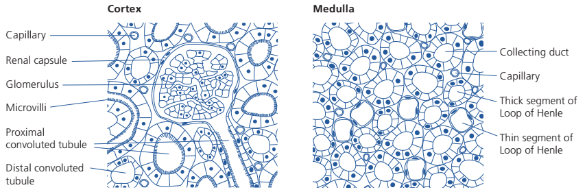
- Found in the cortex, it is a small, dense capillary network.
- Surrounded by Bowman’s capsule, it will often appear as a cluster of small circular spaces in section views.
- Bowman’s Capsule (Renal Capsule):
- Surrounds the glomerulus in the cortex.
- Look for a thin, cup-like structure around the glomerulus, appearing as a circular or oval shape in cross-sections.
- Proximal Convoluted Tubule (PCT):
- Located in the cortex, close to the glomerulus.
- Identifying Feature: Microvilli on the luminal surface, giving it a fuzzy or dense appearance in micrographs.
- Cuboidal epithelial cells in the PCT are packed with mitochondria, though mitochondria may not always be visible in all images.

- Loop of Henle:
- Extends from the cortex into the medulla.
- Has distinct segments:
- Thin Segment: Thinner, less densely packed cells, often visible in the descending limb.
- Thick Segment: Thicker with cuboidal cells, often found in the ascending limb.
- Distal Convoluted Tubule (DCT):
- Located in the cortex, near the collecting ducts.
- Distinguishing Feature: Absence of microvilli, giving it a clearer lumen than the proximal tubule.
- Collecting Duct:
- Runs through the medulla.
- Larger diameter than other tubules, often with clearly defined cell borders and no microvilli.
- Appears pale or clear, surrounded by thick walls compared to nearby nephrons.
Tips for Differentiating Between the Cortex and Medulla
- Cortex:
- Contains the glomeruli and Bowman’s capsules.
- Look for the proximal convoluted tubules, identifiable by their microvilli. Only the cortex has microvilli-rich proximal tubules.
- Medulla:
- Contains loops of Henle and collecting ducts.
- Proximal convoluted tubules and glomeruli are absent, which is a quick way to determine if you are in the medulla.
Histological Characteristics to Note
Collecting Ducts:
- Wide, with visible cell borders and a larger lumen.
Microvilli Presence:
- Only proximal convoluted tubules have microvilli on their inner surface, which helps increase surface area for reabsorption.
Cell Density and Thickness:
- Thick Segment of the Loop of Henle: Larger cuboidal cells, compared to the thin segment.
- Thin Segment of the Loop of Henle: Narrower lumen, lined with squamous cells.