1.05 Electromagnetic Spectrum
Basics of the Electromagnetic Spectrum
- Definition: The electromagnetic spectrum includes all types of electromagnetic waves, which vary in wavelength and frequency.
- Visible Light: Only a small portion of the electromagnetic spectrum; visible wavelengths range from:
- 400 nm (violet light, shorter wavelength, higher energy) to 700 nm (red light, longer wavelength, lower energy).
- The human brain interprets these varying wavelengths as colors, though color itself is perceived by the brain.

Wavelength, Frequency, and Energy
- Wavelength: The distance between successive wave peaks.
- Shorter wavelengths correspond to higher energy and higher frequency.
- Longer wavelengths have lower energy and lower frequency.
- Frequency: The rate at which waves pass a given point. Since all waves travel at the same speed, shorter wavelengths pass more frequently (higher frequency) than longer wavelengths.

Link Between Wavelength and Resolution
- Resolution in Microscopy: The limit of resolution is influenced by the wavelength of the radiation used to observe a specimen.
- The general rule is that resolution is about half the wavelength of the radiation. Thus, smaller wavelengths provide higher resolution.
- Resolution Limit of Light Microscopy:
- Since visible light’s shortest wavelength is about 400 nm (violet light), the resolution limit of a light microscope is around 200 nm.
- This limit means objects smaller than 200 nm, such as ribosomes (~25 nm), cannot be seen with a light microscope.
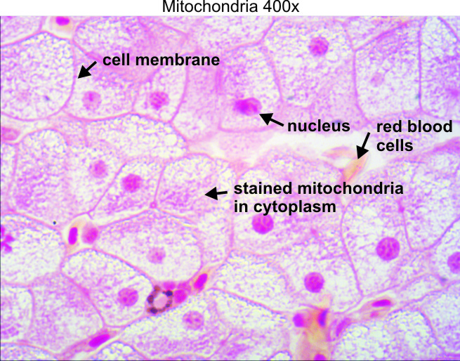
Example: Viewing Mitochondria and Ribosomes
- Mitochondria: Large enough (~0.5-1 μm) to interact with light waves and therefore can be resolved under a light microscope.
- Ribosomes: Much smaller (~25 nm) than half the wavelength of visible light, so they cannot be resolved with a light microscope and remain invisible.
Practical Implications for Microscopy
- Allow visualization of smaller structures like ribosomes that are beyond the resolving power of light microscopes.
- The best resolution achievable with a light microscope is around 200 nm, allowing a maximum useful magnification of about 1500x.
Electron Microscopes:
- Use electron beams with much shorter wavelengths than visible light, offering a much higher resolution.
Practise Questions
1. Multiple Choice
Which of the following best defines the electromagnetic spectrum?
A) The range of wavelengths of visible light only.
B) All types of electromagnetic waves, varying in wavelength and frequency.
C) The spectrum of colors perceived by the human eye.
D) Waves that require a medium to travel.
Answer:
B) All types of electromagnetic waves, varying in wavelength and frequency.
2. Short Answer
Define wavelength and explain its relationship with frequency and energy in electromagnetic waves.
Answer:
Wavelength is the distance between successive wave peaks in an electromagnetic wave. Shorter wavelengths correspond to higher frequencies and higher energy, while longer wavelengths are associated with lower frequencies and lower energy. Since all electromagnetic waves travel at the same speed, shorter wavelengths pass a given point more frequently than longer wavelengths.
3. True or False
True or False: Visible light comprises the majority of the electromagnetic spectrum.
Answer:
False.
Explanation: Visible light is only a small portion of the electromagnetic spectrum, ranging from 400 nm (violet) to 700 nm (red).
4. Calculation-Based Question
If a light microscope uses violet light with a wavelength of 400 nm, what is the approximate resolution limit of this microscope?
Answer:
200 nm
Explanation: The resolution limit is about half the wavelength of the radiation used.
Resolution Limit = 400nm/2 = 200 nm
5. Short Answer
Explain why ribosomes cannot be resolved with a light microscope based on their size and the resolution limit of light microscopy.
Answer:
Ribosomes are approximately 25 nm in size, which is much smaller than half the wavelength of visible light (200 nm). Since the resolution limit of a light microscope is around 200 nm, ribosomes fall well below this threshold and cannot be distinguished as separate entities, rendering them invisible under light microscopy.
6. Multiple Choice
Which statement correctly describes the relationship between wavelength and resolution in microscopy?
A) Longer wavelengths provide higher resolution.
B) Shorter wavelengths provide higher resolution.
C) Wavelength does not affect resolution.
D) Both short and long wavelengths provide the same resolution.
Answer:
B) Shorter wavelengths provide higher resolution.
7. Short Answer
Describe the importance of magnification and resolution in the context of observing cell structures under a microscope.
Answer:
Magnification enables the observation of structures that are too small to be seen with the naked eye by enlarging the image of the object. Resolution determines the level of detail and clarity in the enlarged image, allowing differentiation between closely spaced structures. Together, magnification and resolution are essential for studying cell morphology, organelles, and microorganisms accurately. High magnification without adequate resolution results in blurry images, while high resolution ensures that the magnified image remains clear and detailed.
8. True or False
True or False: Electron microscopes have a lower resolution compared to light microscopes.
Answer:
False.
Explanation: Electron microscopes use electron beams with much shorter wavelengths than visible light, offering a much higher resolution and allowing the observation of finer cellular details.
9. Short Answer
List and briefly describe the two types of electron microscopes mentioned, including their maximum magnification and resolution.
Answer:
- Transmission Electron Microscope (TEM):
- Maximum Magnification: Up to 2,000,000x
- Resolution: About 0.1 nanometers (nm)
- Use: Observing internal structures of cells, viruses, and molecules.
- Scanning Electron Microscope (SEM):
- Maximum Magnification: Up to 500,000x
- Resolution: Approximately 1 nm
- Use: Viewing surface structures in three dimensions, such as cell surfaces and microorganisms.
10. Essay Question
Discuss the relationship between wavelength, frequency, and energy in electromagnetic waves and explain how these properties influence the resolution and magnification capabilities of different types of microscopes.
Answer:
Wavelength, frequency, and energy are fundamental properties of electromagnetic waves that are interrelated. Wavelength is the distance between successive wave peaks, and frequency is the rate at which these waves pass a given point. Shorter wavelengths correspond to higher frequencies and higher energy, while longer wavelengths have lower frequencies and lower energy.
In microscopy, these properties significantly influence resolution and magnification:
- Resolution:
Resolution is the microscope’s ability to distinguish two closely spaced objects as separate. According to the general rule, resolution is about half the wavelength of the radiation used. Therefore, shorter wavelengths (which have higher frequencies and energy) provide higher resolution. This is why electron microscopes, which use electron beams with much shorter wavelengths than visible light, can achieve much higher resolutions (up to 0.1 nm with TEM) compared to light microscopes (with a resolution limit of around 200 nm). - Magnification:
Magnification is the ratio of the size of the image produced by the microscope to the actual size of the object. While magnification itself can be increased by using lenses with higher magnification powers, the useful magnification is limited by the microscope’s resolution. Beyond a certain point, increasing magnification without corresponding resolution leads to image distortion and blurriness. For example, a light microscope can magnify up to 1000x, but its resolution typically limits clear distinction to around 200x. Beyond this, additional magnification does not enhance image quality because the resolution cannot distinguish finer details.
Practical Implications:
Electron Microscopes:
Offer superior resolution, allowing detailed visualization of subcellular structures and microorganisms. The shorter wavelengths of electrons enable the observation of structures at the nanometer scale, which are invisible under light microscopy. However, electron microscopes are expensive, require specialized training, and are unsuitable for observing living cells as specimens must be prepared in a vacuum.
Light Microscopes:
Suitable for observing living cells and tissues due to their ability to work with light and relatively inexpensive setup. However, their resolution is limited by the wavelength of visible light, making it difficult to observe very small structures like ribosomes.