1.04 Units of Measurement and Organelle Sizes
Importance of Small Units in Microscopy
- In microscopy, measuring cellular structures requires extremely small units.
- The International System of Units (SI units) is used worldwide, with the basic length unit being the metre (m).
Metric Prefixes and Conversions
- SI units use standard prefixes to denote multiples or fractions of a base unit.
- Example: kilo- (1000 times), so 1 kilometre = 1000 metres.
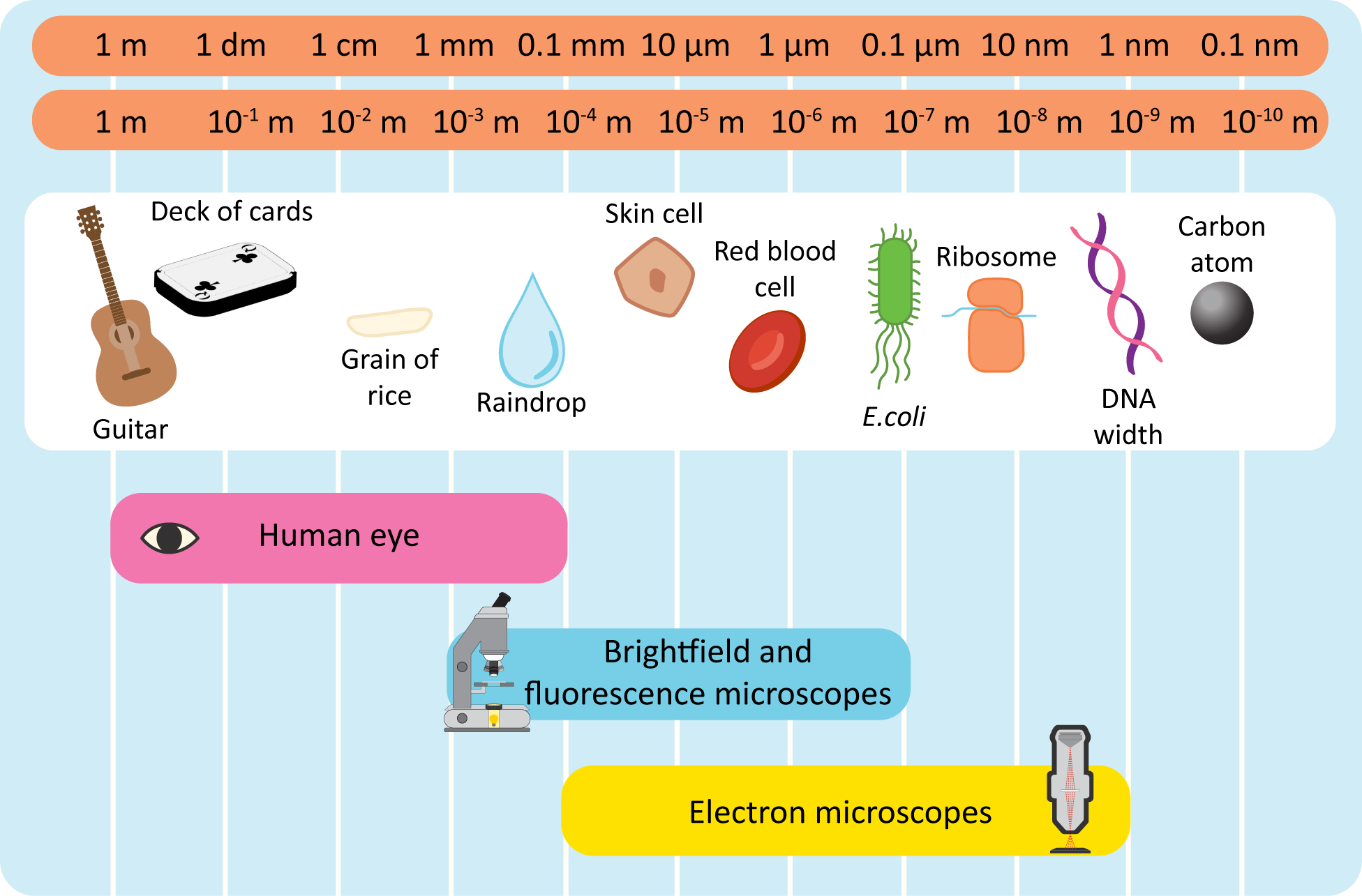
Units of Length Relevant to Cell Studies
- Millimetre (mm): One thousandth of a metre (10⁻³ m).
- Micrometre (μm): One millionth of a metre (10⁻⁶ m); a thousandth of a millimetre.
- Nanometre (nm): One billionth of a metre (10⁻⁹ m); a thousandth of a micrometre.
Organelle Sizes and Visibility Under a Light Microscope
Cell Wall
- Size: 1 – 10 μm (varies with cell type)
- Present In: Plant Cells, Fungi, Some Protists
- Visibility: Yes. Appears as a rigid, outer layer surrounding the cell membrane in plant cells. Typically stained (e.g., with iodine) to enhance visibility under a bright-field microscope.
Vacuole
- Size: 5 – 30 μm
- Present In: Primarily Plant Cells; small in Animal Cells
- Visibility: Yes. In plant cells, a large central vacuole is often visible as a clear or lightly stained area occupying a significant portion of the cell. In animal cells, smaller vacuoles are less prominent and may require specific staining.
Nucleus
- Size: 6 – 10 μm
- Present In: Both Plant and Animal Cells
- Visibility: Yes. The nucleus is usually the most prominent organelle, visible as a large, round or oval structure with a darker region (nucleolus) when stained with dyes like hematoxylin.
Chloroplast
- Size: 4 – 10 μm
- Present In: Plant Cells
- Visibility: Yes. Chloroplasts are visible as green, lens-shaped structures due to the presence of chlorophyll. They can be seen in high concentrations in leaf cells under a light microscope, especially when stained with specific dyes.
Flagella/Cilia
- Size: 5 – 20 μm (Flagella), ~0.2 μm diameter
- Present In: Both Plant and Animal Cells (Flagella in some plant cells like sperm cells; Cilia primarily in animal cells)
- Visibility: Yes (Flagella). Flagella appear as long, whip-like appendages moving in a wave-like manner. Cilia are shorter, more numerous, and beat in coordinated patterns, primarily observed in animal cells like epithelial cells lining the respiratory tract.
Golgi Apparatus
- Size: 1 – 2 μm
- Present In: Both Plant and Animal Cells
- Visibility: Sometimes. When stained, the Golgi apparatus may appear as a series of stacked, flattened sacs (cisternae) near the nucleus, but it can be challenging to distinguish without specific markers.
Endoplasmic Reticulum (ER)
- Size: 0.1 – 1 μm (individual tubules and sheets)
- Present In: Both Plant and Animal Cells
- Visibility: Sometimes. Rough ER can appear as a network of interconnected tubules with ribosomes (giving a “rough” appearance), while Smooth ER lacks ribosomes and appears smoother. Visibility depends on the cell type and staining methods.
Mitochondria
- Size: 0.5 – 1 μm
- Present In: Both Plant and Animal Cells
- Visibility: Rarely. Typically not visible under standard light microscopes due to their small size and lack of distinctive staining. Electron microscopy is required for clear visualization.
Centrosome/Centriole
- Size: 0.5 – 1 μm
- Present In: Animal Cells
- Visibility: Rarely. Difficult to visualize under light microscopy. May appear as small, dot-like structures during specific cell cycle stages if stained with appropriate markers.
Lysosome
- Size: 0.1 – 1.2 μm
- Present In: Animal Cells
- Visibility: No. Generally not visible under light microscopes. Function in breaking down waste materials and cellular debris.
Ribosome
- Size: 0.025 μm (25 nm)
- Present In: Both Plant and Animal Cells
- Visibility: No. Non-membrane-bound organelles responsible for protein synthesis. Visible only with electron microscopy or specialized fluorescence techniques.
Cytoplasm
- Size: Variable (entire cell excluding nucleus and organelles)
- Present In: Both Plant and Animal Cells
- Visibility: Yes. The cytoplasm appears as a gel-like substance filling the cell, providing a medium for organelles to reside and facilitating cellular processes. Organelles are suspended within the cytoplasm and can be seen as distinct structures when stained.
Cytoskeleton
- Size: Variable (fibers range from nanometers to micrometers)
- Present In: Both Plant and Animal Cells
- Visibility: No under standard light microscopy. Composed of microtubules, actin filaments, and intermediate filaments, which provide structural support and facilitate movement within the cell. Visible with fluorescence microscopy using specific dyes.
Microvilli
- Size: 0.1 – 1 μm in diameter; up to 1-2 μm in length
- Present In: Animal Cells (especially epithelial cells)
- Visibility: No under standard light microscopy. These finger-like projections increase surface area for absorption and secretion, particularly in intestinal and kidney cells. Visible with electron microscopy.
Plasmodesmata
- Size: <1 μm
- Present In: Plant Cells
- Visibility: No under light microscopy. Microscopic channels that traverse the cell walls of plant cells, facilitating transport and communication between them. Visible with electron microscopy.
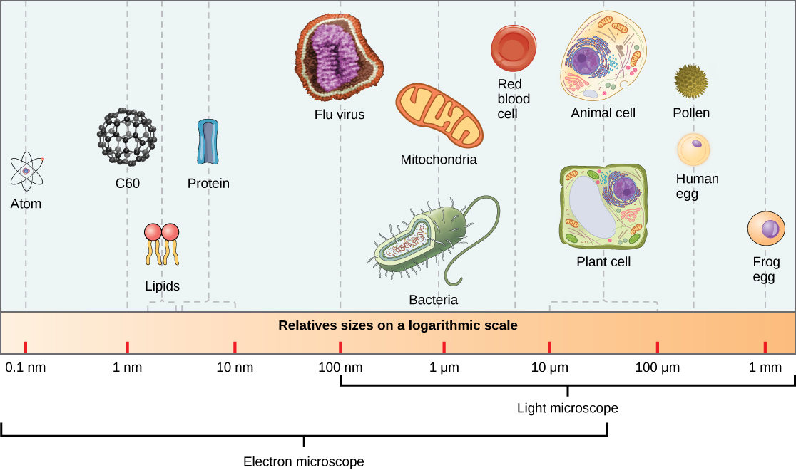
Table of Relevant Units:
| Fraction of a Metre | Unit | Symbol |
|---|---|---|
| One thousandth (10⁻³) | millimetre | mm |
| One millionth (10⁻⁶) | micrometre | μm |
| One billionth (10⁻⁹) | nanometre | nm |
Note: μ\muμ represents the Greek letter “mu”; 1 micrometre = 1/1000 of a millimetre, and 1 nanometre = 1/1000 of a micrometre.
Conversion Examples
| Conversion | Conversion Factor | Example Calculation | Result |
|---|
| Millimeters (mm) to Micrometers (μm) | 1 mm = 1,000 μm | 3 mm × 1,000 | 3,000 μm |
| Centimeters (cm) to Millimeters (mm) | 1 cm = 10 mm | 4.2 cm × 10 | 42 mm |
| Millimeters (mm) to Nanometers (nm) | 1 mm = 1,000,000 nm | 0.02 mm × 1,000,000 | 20,000 nm |
| Centimeters (cm) to Micrometers (μm) | 1 cm = 10,000 μm | 1.5 cm × 10,000 | 15,000 μm |
| Millimeters (mm) to Centimeters (cm) | 10 mm = 1 cm | 25 mm ÷ 10 | 2.5 cm |
Practise Questions
1. Multiple Choice
Which of the following units is the smallest and most appropriate for measuring the size of ribosomes?
A) Millimetre (mm)
B) Micrometre (μm)
C) Nanometre (nm)
D) Centimetre (cm)
Answer: C) Nanometre (nm)
Reason: Ribosomes are extremely small cellular structures, typically measuring around 25 nanometres (nm) in diameter.
2. Question
For each organelle listed below, indicate whether it is visible under a standard light microscope and provide its approximate size range:
- Nucleus
- Ribosome
- Chloroplast
- Mitochondria
- Lysosome
Answer:
Nucleus
- Visibility: Yes
- Size: 6 – 10 μm
Ribosome
- Visibility: No
- Size: 0.025 μm (25 nm)
Chloroplast
- Visibility: Yes
- Size: 4 – 10 μm
Mitochondria
- Visibility: Rarely
- Size: 0.5 – 1 μm
Lysosome
- Visibility: No
- Size: 0.1 – 1.2 μm
4. Comparison
Compare the visibility of the Golgi apparatus and the endoplasmic reticulum (ER) under a standard light microscope.
Answer:
Both the Golgi apparatus and the endoplasmic reticulum (ER) are sometimes visible under a standard light microscope, primarily when stained with specific dyes. The Golgi apparatus may appear as a series of stacked, flattened sacs (cisternae) near the nucleus, but it is challenging to distinguish without specific markers. The Rough ER can sometimes be seen as a network of interconnected tubules with ribosomes, giving it a “rough” appearance, while the Smooth ER appears smoother due to the absence of ribosomes. Visibility depends on the cell type and the staining method used.
5. Application
A biologist measures a plant cell to be 15 μm in diameter. Convert this measurement to nanometres (nm).
Answer:
To convert micrometres (μm) to nanometres (nm):
1 μm = 1,000 nm
15 μm × 1,000 nm/μm = 15,000 nm
Result:
The plant cell is 15,000 nm in diameter.
6. Which organelle is typically not visible under a standard light microscope due to its small size and lack of distinctive staining?
A) Nucleus
B) Chloroplast
C) Mitochondria
D) Vacuole
Answer: C) Mitochondria
7. Application
A plant cell has a large central vacuole measuring 25 μm in diameter. Convert this measurement to millimetres (mm).
Answer:
1 μm = 0.001 mm
25 μm × 0.001 mm/μm = 0.025 mm
Result:
The central vacuole is 0.025 mm in diameter.