1.02 Animal and Plant Cells and Organelles

Organelles
- Organelles are specialized, membrane-bound structures within a cell that perform distinct and essential functions necessary for the cell’s survival, growth, and overall operation.
- Often compared to the organs of a multicellular organism, each organelle has a specific role that contributes to the cell’s health and efficiency.
Cell Structure and Compartmentalization
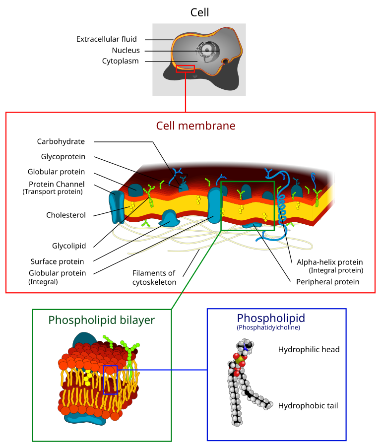
Cell Surface Membrane (Plasma Membrane)
Definition
- A selectively permeable membrane (allows specific molecules/ions to pass) found in plant and animal cells, regulating exchange with the environment.
Structure
- Phospholipid bilayer (double layer of phospholipids):
- Hydrophilic heads (water-attracting) face aqueous surroundings.
- Hydrophobic tails (water-repelling) face inward.
- Trilaminar appearance (three-layered look under EM).
- Includes proteins, cholesterol, and glycoproteins (proteins with sugar chains).
Function
- Barrier: Separates cytoplasm from external surroundings.
- Selective transport: Regulates entry/exit of substances.
- Signalling: Receptors detect external signals (e.g., hormones).
- Recognition: Surface antigens identify cell types.
- Adhesion: Binds cells together for tissue formation.
Role
- Maintains homeostasis (internal balance) by controlling the movement of substances like nutrients, ions, and waste.
- Facilitates communication between cells through signalling pathways.
Key Features
- 7 nm thickness: Very thin for efficient exchange.
- Fluid mosaic model (dynamic structure of lipids and proteins).
- Anchors cytoskeleton (internal cell framework).
- Stabilized by hydrogen bonds with water molecules.
Cytoplasm
Definition
- The gel-like substance within the cell membrane that contains organelles and facilitates cellular processes.
Structure
- Composed of:
- Cytosol (fluid portion): Aqueous solution with ions, nutrients, and enzymes.
- Organelles: Structures like mitochondria, ribosomes, etc., suspended in the cytosol.
- Cytoskeleton: Network of protein fibers (provides structure and transport).
Function
- Medium for biochemical reactions: Site of key metabolic activities (e.g., glycolysis).
- Support: Maintains cell shape and positions organelles.
- Transport: Facilitates intracellular transport of molecules and organelles.
- Storage: Reservoir for nutrients, ions, and waste products.
Role
- Acts as the site of metabolism and supports cellular activity.
- Links organelles, enabling communication and coordination.
Key Features
- Semi-fluid consistency (viscous but dynamic).
- Contains 70-90% water with dissolved substances.
- Dynamic structure with continuous movement of organelles and molecules.


Nucleus & Nucleolus
Nucleus
Definition:
- A membrane-bound organelle (surrounded by a double membrane) that contains the cell’s genetic material (DNA).
Structure:
- Nuclear Envelope: Double membrane with nuclear pores (protein-lined openings) for material exchange.
- Chromatin: DNA and associated proteins in a loose form (condenses into chromosomes during division).
- Nucleoplasm: Semi-fluid matrix inside the nucleus.
Function:
- Genetic storage: Houses DNA (deoxyribonucleic acid).
- Control center: Regulates gene expression and cellular activities.
- Replication: Site of DNA duplication before cell division.
- RNA synthesis: Produces messenger RNA (mRNA) for protein synthesis.
Key Features:
- Found in eukaryotic cells only.
- Contains chromosomes (linear DNA with histones).

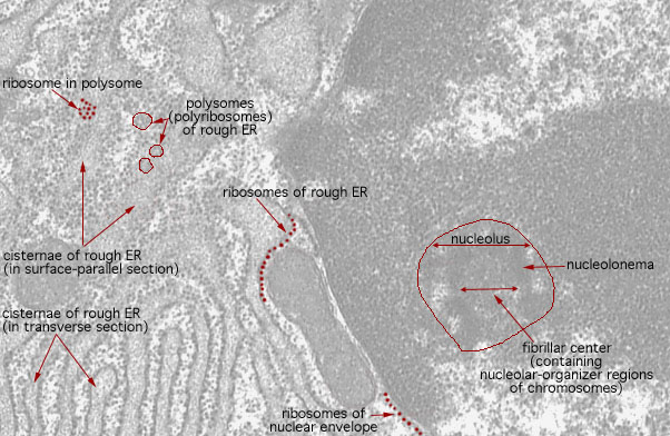
Nucleolus
Definition:
- A dense region within the nucleus where ribosomal RNA (rRNA) is synthesized and ribosome assembly begins.
Structure:
- Comprised of rRNA and proteins.
- No surrounding membrane.
Function:
- Ribosome production: Synthesizes and assembles ribosomal subunits.
- RNA synthesis: Produces rRNA.
- Stress response: Alters activity under cellular stress.
Key Features:
- Visible as a dark-staining body under the microscope.
- Size varies with cell activity (larger in cells with high protein synthesis).

Mitochondria
Definition
- Double-membraned organelle (outer and inner membranes) responsible for aerobic respiration and ATP (adenosine triphosphate) production, known as the “powerhouse of the cell.”
Structure
- Outer membrane:
- Smooth and permeable to small molecules.
- Inner membrane:
- Folded into cristae (increases surface area for reactions).
- Contains enzymes and proteins for the electron transport chain.
- Matrix:
- Gel-like substance inside the inner membrane, contains enzymes for the Krebs cycle, mitochondrial DNA, and ribosomes.
- Intermembrane space:
- Space between the two membranes, involved in proton gradient formation for ATP synthesis.
Function
- ATP production: Generates energy via oxidative phosphorylation.
- Metabolism: Site of the Krebs cycle and electron transport chain.
- Calcium storage: Regulates intracellular calcium levels.
- Heat production: Involved in thermogenesis.
- Apoptosis regulation: Plays a role in programmed cell death.
Role
- Central to energy metabolism and maintaining the cell’s energy supply.
Key Features
- Self-replicating with its own circular DNA and ribosomes.
- Found in high numbers in energy-demanding cells (e.g., muscle, liver).
- Dynamic: Constantly changing shape and location within the cell.


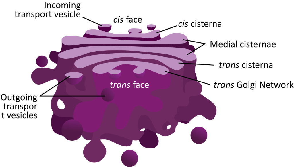
Golgi Apparatus
Definition
- A membrane-bound organelle in eukaryotic cells that processes, modifies, and packages proteins and lipids for secretion or internal use.
Structure
- Cisternae:
- Stacked, flattened membrane sacs.
- Cis face: Receives vesicles from the ER.
- Trans face: Sends out processed vesicles to their destinations.
- Vesicles:
- Membrane-bound sacs for transport to and from the Golgi.
Function
- Modification: Adds carbohydrates (glycosylation) and phosphates to proteins and lipids.
- Packaging: Sorts materials into vesicles for transport.
- Lysosome formation: Produces and packages digestive enzymes.
- Secretion: Prepares materials for export (e.g., hormones, enzymes).
- Membrane renewal: Supplies components for the cell membrane.
Role
- Central in the secretory pathway and post-translational modification of biomolecules.
Key Features
- Dynamic structure: Constantly forming and breaking down vesicles.
- Works closely with the endoplasmic reticulum (ER) for material processing and transport.
- Prominent in secretory cells (e.g., pancreatic or salivary gland cells).

Endoplasmic Reticulum (ER)
Definition
- A network of membrane-bound tubules and sacs (cisternae) involved in protein and lipid synthesis, transport, and storage in eukaryotic cells.
Types
- Rough ER (RER):
- Studded with ribosomes (protein-synthesizing structures).
- Involved in protein synthesis and transport to the Golgi.
- Smooth ER (SER):
- Lacks ribosomes.
- Synthesizes lipids, detoxifies chemicals, and regulates calcium levels.
Structure
- Continuous with the nuclear envelope (double membrane of the nucleus).
- Composed of interconnected cisternae, tubules, and vesicles.
Function
- Rough ER:
- Synthesizes and transports proteins for secretion or membrane insertion.
- Smooth ER:
- Lipid and steroid hormone synthesis.
- Detoxification of drugs and toxins (especially in liver cells).
- Calcium storage and release in muscle cells.
Role
- Central in protein and lipid metabolism.
- Acts as an intracellular transport system for biomolecules.
Key Features
- Abundant in cells with high protein production (e.g., pancreatic cells) or lipid metabolism (e.g., liver cells).
- Works closely with the Golgi Apparatus and ribosomes for cellular function.
- Provides compartmentalization within the cell for efficient biochemical processes.


Ribosomes
Definition
- Non-membrane-bound organelles composed of RNA and proteins that are responsible for protein synthesis in cells.
Structure
- Subunits:
- Large subunit: Catalyzes peptide bond formation.
- Small subunit: Binds to mRNA for translation.
- Composition:
- Made of ribosomal RNA (rRNA) and proteins.
- Types:
- 80S ribosomes: Found in eukaryotic cells (larger).
- 70S ribosomes: Found in prokaryotic cells, mitochondria, and chloroplasts (smaller).
Function
- Translation:
- Assembles amino acids into polypeptides based on the mRNA sequence.
- Protein synthesis: Produces proteins for intracellular use (cytoplasmic ribosomes) or export (ribosomes on RER).
Role
- Essential for gene expression by converting genetic information into functional proteins.
Location:
- Free ribosomes in the cytoplasm produce intracellular proteins.
- Bound ribosomes on the rough ER synthesize proteins for secretion or membrane integration.
- Found in both prokaryotic and eukaryotic cells.
- Consists of ribonucleoproteins (RNA + protein complex).

Lysosomes
Definition
- Membrane-bound organelles containing digestive enzymes that break down biological molecules, cellular debris, and foreign material.
Structure
- Single membrane:
- Protects the cell from the enzymes inside.
- Acidic interior (pH ~5):
- Optimal for enzyme activity.
- Contains hydrolytic enzymes (e.g., proteases, lipases, nucleases).
Function
- Intracellular digestion: Breaks down macromolecules, damaged organelles (autophagy), and pathogens (phagocytosis).
- Waste removal: Processes cellular waste for recycling or excretion.
- Apoptosis: Releases enzymes to aid in programmed cell death.
- Membrane repair: Participates in repairing damaged cell membranes.
Role
- Central to cellular recycling and maintaining homeostasis by removing unwanted materials.
Key Features
- Found in eukaryotic cells (particularly in animal cells).
- Derived from the Golgi apparatus.
- Active in immune cells (e.g., macrophages).
- Defects in lysosome function cause diseases (e.g., Tay-Sachs disease).

Centrioles (Animal Cells Only)
Definition
- Cylindrical structures made of microtubules that play a role in cell division and organizing the cytoskeleton.
Structure
- Microtubules:
- Composed of nine triplets arranged in a cylindrical pattern (9+0 arrangement).
- Found in pairs, called a centrosome when located near the nucleus.
Function
- Spindle fiber formation:
- Organize and anchor microtubules during mitosis/meiosis to ensure proper chromosome separation.
- Cytoskeleton organization:
- Help maintain cell shape and structure.
- Cilia and flagella formation:
- Act as basal bodies for the assembly of cilia and flagella.
Role
- Essential in cell division and intracellular organization in animal cells.
Key Features
- Present only in animal cells.
- Duplicate during the cell cycle to ensure each daughter cell has one pair.
- Absent in most plant cells, fungi, and prokaryotes.

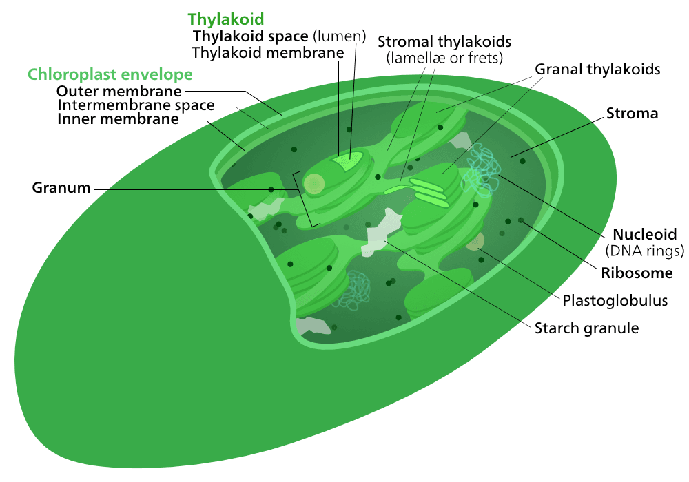
Chloroplasts (Plant Cells Only)
Definition
- Double-membraned organelles in plant cells responsible for photosynthesis and converting light energy into chemical energy (glucose).
Structure
- Outer membrane: Smooth and permeable.
- Inner membrane: Regulates materials entering and exiting the chloroplast.
- Stroma: Fluid-filled interior containing enzymes, DNA, and ribosomes.
- Thylakoids: Flattened sacs containing chlorophyll (light-absorbing pigment).
- Arranged in stacks called grana.
- Linked by lamellae (membrane bridges).
- Chlorophyll: Green pigment crucial for light absorption.
Function
- Photosynthesis: Converts sunlight into glucose and oxygen:
- Light-dependent reactions: Occur in thylakoid membranes.
- Light-independent reactions (Calvin cycle): Occur in the stroma.
- Storage: Temporary storage of starch.
- Energy production: Provides chemical energy in the form of ATP and NADPH.
Role
- Primary site of energy production in plants.
- Maintains oxygen levels through oxygen release during photosynthesis.
Key Features
- Found in plant cells and some algae.
- Contains circular DNA and ribosomes, allowing it to self-replicate.
- Absent in animal cells.


Cell Wall (Not found in animal cells)
Definition:
- A rigid, extracellular structure made primarily of cellulose that provides support and protection to plant cells.
Structure:
- Primary Cell Wall: Flexible and thin, composed of cellulose microfibrils.
- Secondary Cell Wall: Thicker, contains lignin for additional strength (in some cells).
- Middle Lamella: Pectin-rich layer between adjacent cells for adhesion.
Function:
- Support: Maintains cell shape and prevents bursting from osmotic pressure.
- Protection: Acts as a barrier against pathogens.
- Permeability: Allows water, ions, and small molecules to pass freely.
- Communication: Works with plasmodesmata for intercellular transport.
Role:
- Essential for structural integrity and osmotic balance in plants.
Key Features:
- Found in plant cells, fungi, and some prokaryotes (composition differs).
- Composed of cellulose, hemicellulose, and pectin in plants.

Plasmodesmata
Definition:
- Microscopic channels through the cell wall that connect adjacent plant cells, facilitating intercellular transport and communication.
Structure:
- Lined by the plasma membrane and contain a desmotubule (ER-derived structure) within the channel.
Function:
- Transport: Allows passage of water, ions, and small molecules between cells.
- Communication: Enables direct cell-to-cell signaling.
- Coordination: Helps maintain tissue and organ function by synchronizing cellular activities.
Role:
- Crucial for symplastic transport (movement through cytoplasm) and plant development.
Key Features:
- Unique to plant cells.
- Allows integration of cellular activities across a tissue.

Vacuoles
Definition
- Membrane-bound organelles that store nutrients, waste, and water, and maintain turgor pressure in plant cells.
Structure
- Tonoplast:
- A selectively permeable membrane surrounding the vacuole.
- Content:
- Contains water, ions, enzymes, waste products, and pigments.
- Size:
- Large central vacuole in plant cells; smaller, temporary vacuoles in animal cells.
Function
- Storage: Reserves water, nutrients, and waste products.
- Turgor Pressure: Maintains rigidity by exerting pressure on the cell wall.
- Waste Management: Isolates harmful substances.
- pH Balance: Regulates cell sap pH.
- Pigment Storage: Stores pigments (e.g., anthocyanins) for flower/fruit coloration.
Role
- In plant cells, it supports structure and aids in growth.
- In animal cells, it temporarily stores substances or aids in waste removal.
Key Features
- Large central vacuole in plants (dominates cell volume).
- Dynamic: Size and function vary based on the cell’s needs.
- Found in eukaryotic cells (more prominent in plants).

Differences Between Animal and Plant Cells
Unique to Animal Cells
- Centrioles: Involved in organizing microtubules during cell division.
- Vacuoles: Typically small and temporary.
Unique to Plant Cells
- Cell Walls: Provide structural support and protection.
- Chloroplasts: Enable photosynthesis.
- Large Central Vacuole: Maintains cell rigidity and stores substances.
- Plasmodesmata: Allow intercellular communication and transport.
Comparative Summary
| Feature | Animal Cells | Plant Cells |
|---|---|---|
| Centrioles | Present | Absent |
| Cell Wall | Absent | Present (cellulose-based) |
| Chloroplasts | Absent | Present |
| Vacuoles | Small and temporary | Large and permanent central vacuole |
| Plasmodesmata | Absent | Present |
| Shape | Irregular, flexible shapes | Typically fixed, rectangular shapes |
Practise Questions
1. Multiple Choice
Which organelle is unique to animal cells and plays a crucial role in cell division?
A) Chloroplast
B) Centrioles
C) Large central vacuole
D) Cell wall
Answer: B) Centrioles
2. Short Answer
Define compartmentalization in the context of cellular biology and explain its significance in eukaryotic cells.
Answer:
Compartmentalization refers to the division of a cell into distinct regions or organelles, each enclosed by membranes. This allows specific cellular processes to occur in separate environments, enhancing efficiency and organization. In eukaryotic cells, compartmentalization enables complex functions by isolating different biochemical pathways, preventing interference between processes, and allowing specialized environments for activities like protein synthesis in the endoplasmic reticulum or energy production in mitochondria.
3. Diagram-Based Question

Label the following structures on both the plant and animal cell diagrams provided:
- Cell Wall
- Chloroplast
- Large Central Vacuole
- Centrioles
- Nucleus
- Mitochondria
- Golgi Apparatus
- Cytoplasm
- Cell Surface Membrane
- Plasmodesmata
Answer:
Note that:
- Plant Cell: Will have Cell Wall, Chloroplast, Large Central Vacuole, Plasmodesmata.
- Animal Cell: Will have Centrioles (if included), but not Cell Wall, Chloroplast, or Large Central Vacuole.
4. Comparison
Compare the structure and function of vacuoles in plant and animal cells.
Answer:
In plant cells, vacuoles are typically large, permanent central vacuoles surrounded by a tonoplast membrane. They store water, pigments, enzymes, sugars, and other solutes, regulating osmotic balance and maintaining turgor pressure, which provides structural support to the plant. Additionally, they store pigments that give color to flowers and vegetables.
In animal cells, vacuoles are usually small and temporary, such as phagocytic vacuoles involved in engulfing and digesting foreign particles. They play roles in storing nutrients and waste products but do not provide structural support as in plant cells.
5. Application
Explain how the presence of chloroplasts and mitochondria in plant cells supports their metabolic processes.
Answer:
Chloroplasts are essential for photosynthesis, allowing plant cells to convert light energy into chemical energy (glucose). They contain chlorophyll, which absorbs light energy, and structures like grana where the light-dependent reactions occur.
Mitochondria perform aerobic respiration, breaking down glucose to produce ATP, the cell’s main energy source. This dual presence ensures that plant cells can generate energy both through photosynthesis (producing glucose) and respiration (utilizing glucose), supporting their growth, maintenance, and metabolic activities.
6. True or False
True or False: Plasmodesmata are found in both plant and animal cells and facilitate the movement of molecules between cells.
Answer:
False. Plasmodesmata are unique to plant cells and are cytoplasmic strands that pass through the cell walls, allowing the transport and communication between adjacent plant cells. Animal cells do not have plasmodesmata; instead, they use structures like gap junctions for intercellular communication.
7. Short Answer
Describe the role of the Golgi apparatus in a plant cell.
Answer:
The Golgi apparatus in a plant cell functions as the cell’s internal sorting and distribution center. It modifies, sorts, and packages proteins and lipids synthesized in the endoplasmic reticulum for transport to their destinations, either within the cell or for secretion outside the cell. This organelle is essential for processing molecules that are crucial for various cellular functions, including the synthesis of the cell wall components.
8. Explanation
Explain how the structure of the cell wall in plant cells prevents cell bursting during osmosis.
Answer:
The cell wall in plant cells is a rigid, thick layer composed primarily of cellulose fibers. This structure provides strength and rigidity, allowing the cell to withstand high internal pressures that develop when water enters the cell through osmosis. By restricting excessive expansion, the cell wall prevents the cell from bursting, maintaining structural integrity and shape even under significant osmotic pressure.
9. Multiple Choice
Which of the following structures connects plant cells and allows the transport of molecules between them?
A) Centrioles
B) Plasmodesmata
C) Gap Junctions
D) Tight Junctions
Answer: B) Plasmodesmata
10. Question
Discuss the key differences between animal and plant cells, highlighting how these differences relate to their respective functions and lifestyles.
Answer:
Key Differences:
- Cell Wall:
- Plant Cells: Possess a rigid cell wall made of cellulose, providing structural support, maintaining cell shape, and protecting against mechanical stress. This rigidity is essential for maintaining the upright structure of plants.
- Animal Cells: Lack a cell wall, allowing for a more flexible cell shape, which is necessary for the diverse forms and movements in animals.
- Chloroplasts:
- Plant Cells: Contain chloroplasts, enabling photosynthesis to convert light energy into chemical energy (glucose), which supports the plant’s growth and energy needs.
- Animal Cells: Do not have chloroplasts and must obtain energy by consuming other organisms.
- Vacuoles:
- Plant Cells: Have a large, central vacuole that stores water, nutrients, and waste products, and maintains turgor pressure for structural support.
- Animal Cells: Contain small, temporary vacuoles used for storage and transport within the cell but do not contribute significantly to cell rigidity.
- Centrioles:
- Animal Cells: Possess centrioles that play a critical role in cell division by organizing the mitotic spindle.
- Plant Cells: Generally lack centrioles, and cell division is organized differently without these structures.
- Plasmodesmata:
- Plant Cells: Have plasmodesmata, allowing direct communication and transport between adjacent cells, which is vital for coordinating functions in multicellular plants.
- Animal Cells: Use different structures like gap junctions for intercellular communication.
Relation to Functions and Lifestyles:
- Intercellular Communication: Plasmodesmata in plant cells enable synchronized activities across cells, essential for growth, nutrient distribution, and response to environmental changes.
- Structural Support: The presence of a cell wall in plant cells supports their stationary lifestyle, providing the necessary rigidity to remain upright and withstand environmental stresses.
- Energy Acquisition: Chloroplasts enable plants to produce their own food through photosynthesis, allowing them to thrive in various environments without needing to consume other organisms.
- Cell Division: Centrioles in animal cells facilitate the efficient segregation of chromosomes during cell division, which is crucial for the development and maintenance of complex animal tissues.