1.08 Measuring cells
Magnification and Resolution
- Magnification:
- Describes how much larger an image appears compared to the actual size of the object.
- Resolution:
- Resolution is the ability to distinguish two close points as separate. Higher resolution reveals finer details.
Why a Limit Exists:
- The resolution of light microscopes is limited by the wavelength of light, meaning fine details smaller than about 200 nanometers (nm) are indistinguishable.
- Electron microscopes use electron beams with shorter wavelengths than light, providing much higher resolution and allowing scientists to see sub-cellular structures invisible under a light microscope.
- Electron microscopy was developed to overcome this limit, allowing scientists to observe structures at a much smaller scale.
Calculations:
- Observed Size of Image (I): The size of the object as it appears under the microscope (what can be measured with a ruler or scale on the image).
- Actual Size (A): The true size of the object being viewed, before any magnification.
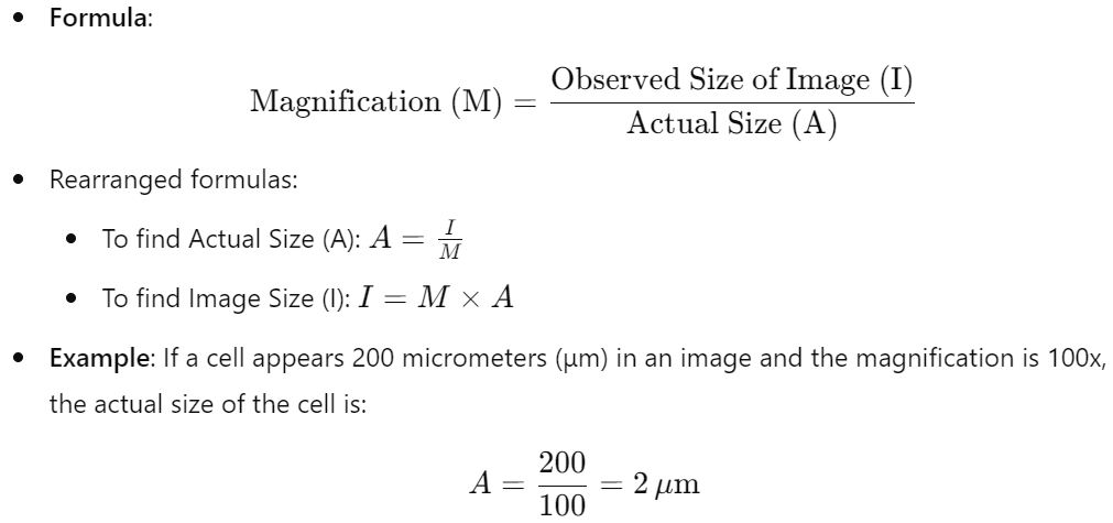
Measuring Cells Using an Eyepiece Graticule
- Eyepiece Graticule:
- A transparent scale inserted into the microscope eyepiece, typically with 100 divisions, used for measuring cells and structures. These divisions in themselves do not have units. We assign units to them with the help of a stage micrometer.
- To measure, align the object with the graticule; the difference between scale points gives a measurement in eyepiece units.
- Calibration of the Eyepiece Graticule:
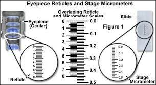
- Stage Micrometer: A microscopic ruler with known measurements:
- 1 mm total length
- Subdivided into 10 larger decisions
- Each of the 10 larger divisions were further subdivided into 10 smaller 0.01 mm for division
- ..is placed on the microscope stage.
- Stage Micrometer: A microscopic ruler with known measurements:
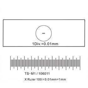
- Align the eyepiece graticule with the stage micrometer to calibrate the value of each eyepiece unit in micrometers (μm).
Worked Example for Measuring Cells
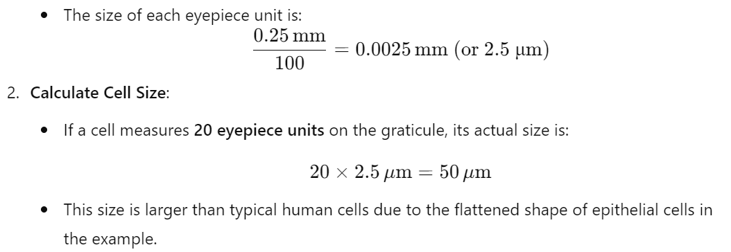
- Suppose 100 eyepiece units align with 0.25 mm on the stage micrometer.
Calibrate the Eyepiece Graticule:
- Suppose 100 eyepiece units align with 0.25 mm on the stage micrometer.
Practise Questions
1. Multiple Choice
Which of the following best defines magnification in microscopy?
A) The ability to distinguish two close points as separate entities.
B) The degree to which an image appears larger than the actual size of the object.
C) The quality of the image produced by the microscope.
D) The wavelength of light used to illuminate the specimen.
Answer:
B) The degree to which an image appears larger than the actual size of the object.
2. Short Answer
Define resolution and explain its significance in microscopy.
Answer:
Resolution is the ability of a microscope to distinguish two closely spaced points as separate and distinct entities. It is significant because higher resolution allows for clearer and more detailed images of the specimen, enabling the observation of finer cellular structures and details that are essential for accurate analysis and understanding in cell biology.
3. True or False
True or False: The resolution of a light microscope is limited by the wavelength of light, making it difficult to distinguish details smaller than approximately 200 nanometers.
Answer:
True. The resolution of light microscopes is indeed limited by the wavelength of visible light, typically around 200 nanometers, which restricts the ability to distinguish finer details smaller than this threshold.
4. Calculation-Based Question
A microscope has an eyepiece lens with a magnification of 10x and an objective lens with a magnification of 40x. Calculate the total magnification of the microscope.
Answer:
Total Magnification = Magnification of Eyepiece Lens × Magnification of Objective Lens
Total Magnification = 10x × 40x = 400x
5. Short Answer
Explain why electron microscopes can achieve higher resolution compared to light microscopes.
Answer:
Electron microscopes can achieve higher resolution than light microscopes because they use electron beams, which have much shorter wavelengths than visible light. The shorter wavelength of electrons allows electron microscopes to distinguish finer details and structures at the nanometer scale, overcoming the resolution limit of approximately 200 nanometers inherent to light microscopes.
6. Short Answer
Describe the purpose of an eyepiece graticule in measuring cells under a microscope.
Answer:
An eyepiece graticule is a transparent scale inserted into the microscope’s eyepiece, typically divided into 100 units. It is used as a measuring tool to determine the size of cells and structures viewed under the microscope. By aligning the object with the graticule and counting the number of divisions it spans, researchers can obtain measurements in eyepiece units, which are then converted to actual size using a calibration factor derived from a stage micrometer.
7. Calculation-Based Question
Suppose 100 eyepiece units align with 0.25 mm on the stage micrometer. Calculate the calibration factor in micrometers (µm) per eyepiece unit.
Answer:
Calibration Factor = Known Distance on Stage Micrometer / Number of Eyepiece Divisions
Calibration Factor = 0.25 mm / 100 divisions = 0.0025 mm/division
Convert mm to micrometers: 0.0025 mm = 2.5 µm
Calibration Factor = 2.5 µm per eyepiece unit
8. Application
A cell spans 30 eyepiece units on the graticule. Using the calibration factor from Question 7, calculate the actual size of the cell in micrometers (µm).
Answer:
Actual Size = Number of Eyepiece Divisions × Calibration Factor
Actual Size = 30 × 2.5 µm = 75 µm
9. Multiple Choice
Which factor primarily limits the resolution of a light microscope?
A) The numerical aperture of the objective lens
B) The wavelength of light used
C) The magnification of the eyepiece
D) The intensity of the light source
Answer:
B) The wavelength of light used
10. Essay Question
Discuss the importance of magnification and resolution in cell biology studies. Include in your discussion the limitations of light microscopes and how electron microscopes address these limitations.
Answer:
Magnification and resolution are fundamental parameters in cell biology that determine the level of detail and clarity observable under a microscope. Magnification allows scientists to enlarge the image of small structures, making it possible to observe cells and their internal components that are otherwise invisible to the naked eye. However, magnification alone does not enhance the clarity of the image; this is where resolution becomes crucial. Resolution is the ability to distinguish two closely spaced points as separate, providing the clarity and detail necessary to study cellular structures accurately.
The limitations of light microscopes stem primarily from the wavelength of visible light, which restricts resolution to about 200 nanometers. This limitation means that structures smaller than this threshold, such as ribosomes, individual proteins, and certain organelles, cannot be clearly distinguished. Additionally, light microscopes often require staining techniques to enhance contrast, which can sometimes alter or obscure cellular structures.
Electron microscopes overcome these limitations by utilizing electron beams, which have much shorter wavelengths than visible light, achieving resolutions up to 0.1 nanometers. This allows for the detailed visualization of intricate sub-cellular structures, including the detailed architecture of organelles like mitochondria, the endoplasmic reticulum, and the Golgi apparatus.