1.06 Electron Microscope
1. Limitations of Light Microscopy
- Resolution Limit:
- Maximum Resolution: ~200 nm
- Reason: Limited by the wavelength of visible light (400-700 nm)
- Implications: Inability to visualize smaller structures such as organelles, viruses, and molecular complexes.
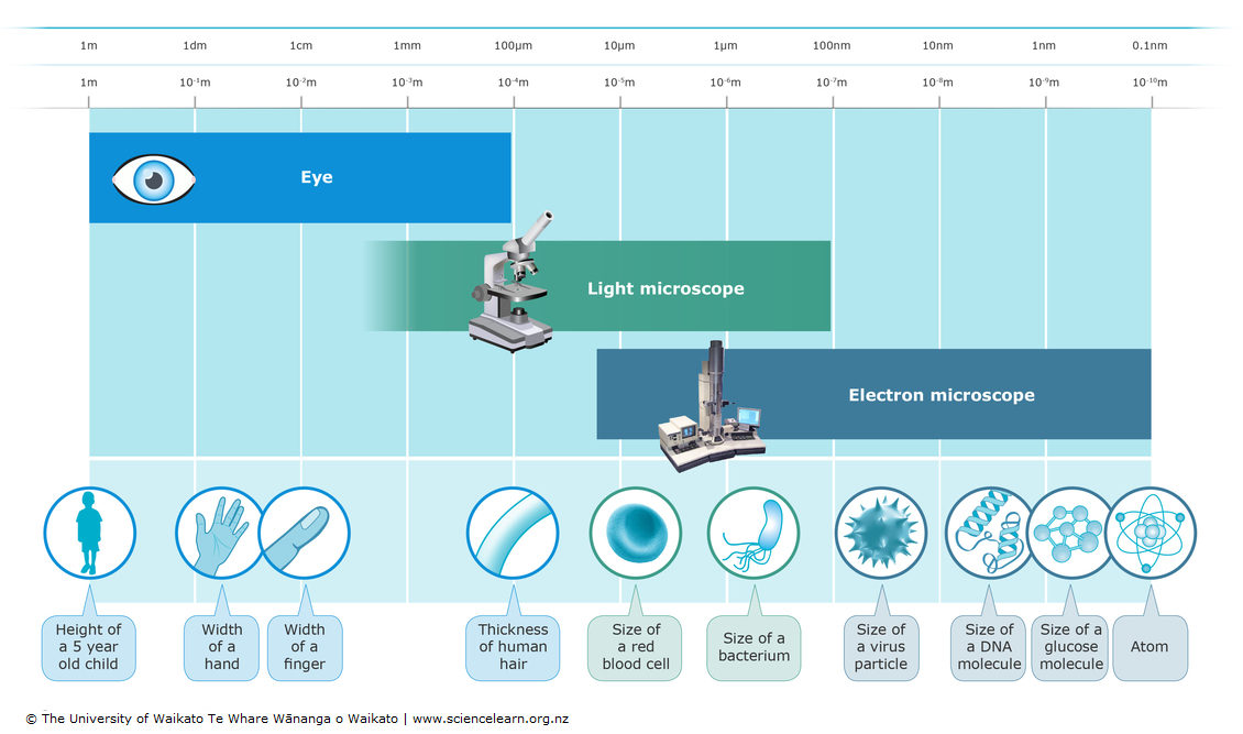
2. Why Use Electrons for Microscopy?
- Short Wavelengths:
- Electrons: ~0.005 nm (varies with energy)
- Advantage: Provides much higher resolution (~0.1-0.5 nm) compared to visible light.
- Electromagnetic Lenses:
- Function: Focus and direct the electron beam with precision, similar to glass lenses in light microscopes.
- Higher Magnification: Enables detailed visualization of structures at the molecular and atomic levels.
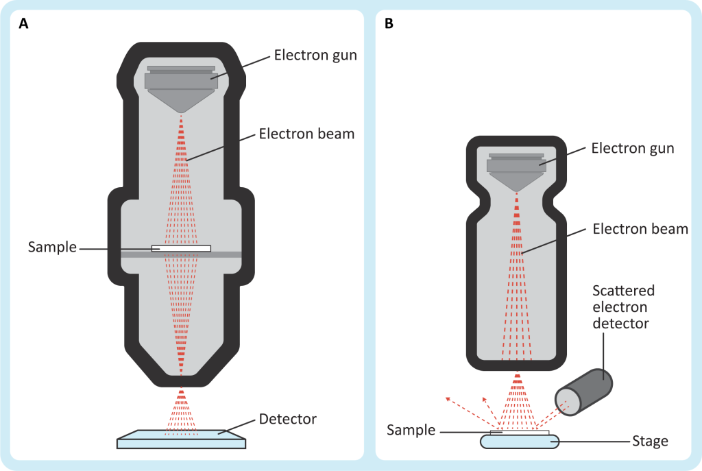
3. How an Electron Microscope (EM) Works
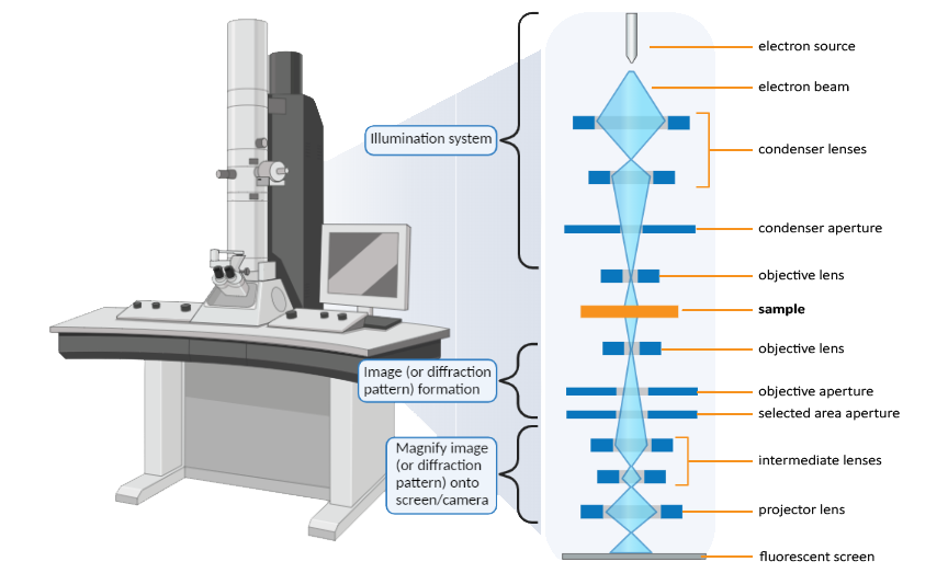
3.1. Components of an Electron Microscope
- Electron Gun and Anode:
- Electron Gun: Generates a beam of electrons by heating a metal filament (thermionic emission).
- Anode: Accelerates electrons into a high-speed, focused beam directed towards the specimen.
- Condenser Electromagnetic Lens:
- Function: Shapes and directs the electron beam onto the specimen.
- Purpose: Ensures even illumination and precise focusing of the electron beam.
- Specimen Placement:
- Support: Specimen is placed on a thin metal grid instead of a glass slide to prevent interference with the electron beam.
- Preparation: Specimens are ultra-thin (especially for Transmission Electron Microscopy, TEM) to allow electrons to pass through.
- Objective Electromagnetic Lens:
- Function: Produces a magnified image of the specimen after interaction with the electron beam.
- Role: Focuses the initial image, which is then further magnified by projector lenses.
- Projector Electromagnetic Lenses:
- Function: Further magnify the image created by the objective lens.
- Output: Projects the final magnified image onto a viewing screen or photographic plate.
- Viewing Screen or Photographic Plate:
- Viewing Screen: Typically a fluorescent screen displaying a monochrome (black and white) image.
- Recording: Images can be captured using photographic plates or digital sensors.
3.2. Additional Processes
- Staining with Heavy Metals:
- Purpose: Enhances contrast by using heavy metals (e.g., osmium tetroxide, lead citrate) that block electrons, creating darker areas in the image.
4. Types of Electron Microscopes
4.1. Transmission Electron Microscope (TEM)
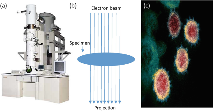
- Function: Examines internal structures within cells by transmitting electrons through ultra-thin specimen sections.
- Resolution: Up to ~0.5 nm.
- Applications: Detailed study of cellular organelles, viruses, and molecular complexes.
4.2. Scanning Electron Microscope (SEM)
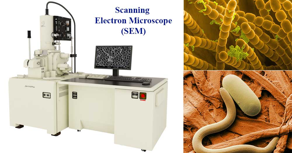
- Function: Scans the surface of specimens with an electron beam, detecting reflected electrons to create three-dimensional images.
- Resolution: Typically between 3 nm and 20 nm.
- Applications: Surface imaging of cells, tissues, microorganisms, and materials science.
5. Comparison: Light Microscopes vs. Electron Microscopes
| Feature | Light Microscope | Electron Microscope |
|---|---|---|
| Source of Radiation | Visible light | Electron beams |
| Wavelength of Radiation | 400-700 nm (visible light spectrum) | ~0.005 nm (varies with electron energy) |
| Maximum Resolution | ~200 nm | 0.1-0.5 nm |
| Lens Type | Glass lenses (convex and concave) | Electromagnetic lenses |
| Specimen State | Can observe live and dead specimens | Typically requires dead specimens (fixed and dehydrated) |
| Staining Agents | Colored dyes (e.g., hematoxylin, eosin) | Heavy metals (e.g., osmium tetroxide, lead citrate) |
| Image Type | Colored photomicrographs | Monochrome (black and white) images |
| Viewing Mechanism | Through an eyepiece or digital display | On a fluorescent screen, digital monitors, or detectors |
| Magnification Range | 40× to 1,000× | 10,000× to 2,000,000× |
| Resolution Limit | Limited by light wavelength (~200 nm) | Higher, limited by electron wavelength (~0.1 nm) |
| Sample Preparation | Minimal; often observed in natural state | Extensive; requires dehydration, fixation, coating with conductive materials |
| Cost | Relatively affordable and widely available | Very expensive, requiring specialized facilities |
| Operating Environment | Standard laboratory settings | Requires vacuum conditions and specialized infrastructure |
| Depth of Field | Greater, allowing viewing of thicker samples | Limited; best for thin, ultra-thin specimens |
| Ease of Use | Easier to use and maintain | Requires specialized training and maintenance |
| Portability | Often portable (e.g., stereo microscopes) | Typically large and non-portable |
| Applications | Viewing cells, tissues, microorganisms, and live specimens | Detailed study of cellular organelles, viruses, nanoparticles, and materials science |
| Speed | Generally faster for routine observations | Slower due to complex sample preparation and imaging process |
| Image Color | Naturally colored (can be enhanced with stains) | Monochromatic (black and white); can be digitally colorized |
6. Specimen Preparation for Electron Microscopy
- Fixation: Preserving the specimen’s structure using chemical fixatives (e.g., glutaraldehyde).
- Dehydration: Removing water through a series of ethanol or acetone washes.
- Embedding: Mounting the specimen in a resin to create thin sections (especially for TEM).
- Sectioning: Cutting ultra-thin slices (50-100 nm) using an ultramicrotome for TEM.
- Staining: Applying heavy metals to enhance contrast.
- Coating (for SEM): Spraying with a conductive material (e.g., gold) to prevent charging under the electron beam.
7. Resolution and Image Quality in Electron Microscopy
- Resolution:
- TEM: Up to ~0.5 nm, allowing visualization of detailed internal structures like ribosomes and viral particles.
- SEM: Typically between 3 nm and 20 nm, suitable for detailed surface topography.
- Image Quality:
- Contrast: Enhanced by heavy metal staining, with denser areas appearing darker.
- Monochromatic Images: Produced in black and white; color can be added digitally if needed.
- Ultrastructure Visualization: Reveals detailed cellular and organelle structures not visible with light microscopy.
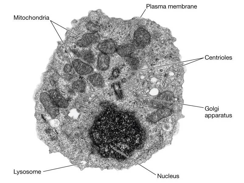
8. Common Plant and Animal Organelles in Electron Micrographs
8.1. Nucleus
- Appearance: Large, prominent structure with a double membrane (nuclear envelope) and visible nuclear pores.
- Features: Contains dense regions called nucleoli.
8.2. Mitochondria
- Appearance: Bean-shaped with a double membrane.
- Features: Inner membrane folded into cristae, appearing as intricate parallel lines to enhance surface area.
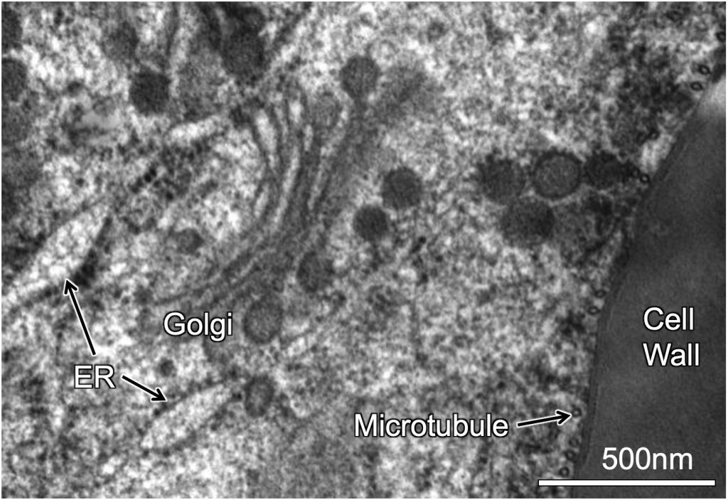
8.3. Endoplasmic Reticulum (ER)
- Rough ER:
- Appearance: Studded with ribosomes as small, dot-like structures.
- Smooth ER:
- Appearance: Lacks ribosomes; appears tubular or sheet-like.
8.4. Golgi Apparatus
- Appearance: Stacked, flattened membrane sacs (cisternae) resembling a series of pancakes.
- Location: Often near the ER; involved in modifying and packaging proteins.
8.5. Ribosomes
- Appearance: Tiny, granular structures; either free in the cytoplasm or attached to the rough ER.
- Appearance: Small dots or specks within the cell.
8.6. Lysosomes
- Appearance: Small, spherical vesicles containing dense, granular material.
- Function: Involved in digestion and waste processing within the cell.
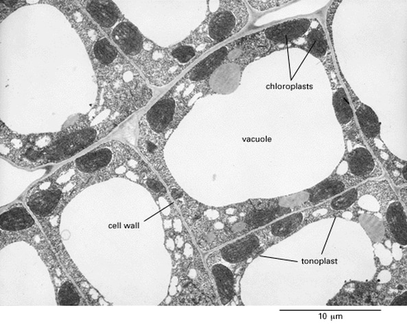
8.7. Chloroplasts (Plant Cells)
- Appearance: Large, disc-shaped organelles with an outer membrane and internal thylakoid membranes arranged in stacks called grana.
- Features: Contain chlorophyll, giving them a distinct appearance.
8.8. Cell Wall (Plant Cells)
- Appearance: Rigid, thick layer outside the cell membrane composed of cellulose fibers.
- Function: Provides structural support and protection.
8.9. Vacuoles (Plant and Animal Cells)
- Plant Cells:
- Appearance: Large central vacuole appears as a clear, empty space.
- Animal Cells:
- Appearance: Smaller, more numerous vacuoles occupying less space.
8.10. Centrosomes and Centrioles (Animal Cells)
- Appearance: Centrosomes contain a pair of centrioles, cylindrical structures arranged perpendicular to each other.
- Function: Play a key role in cell division.
8.11. Cytoskeleton
- Appearance: Network of filamentous structures (microtubules, microfilaments, and intermediate filaments).
- Function: Provides shape and support to the cell; often seen as thin, thread-like structures.
9. Applications of Electron Microscopy
- Biological Research:
- Detailed study of cellular organelles and structures.
- Visualization of viruses and molecular complexes.
- Understanding cellular processes at the ultrastructural level.
- Materials Science:
- Analyzing the surface and internal structure of materials.
- Studying nanoparticles and nanomaterials.
- Medical Diagnostics:
- Identifying pathogens and cellular abnormalities.
- Researching tissue samples for disease diagnosis.
10. Advantages and Limitations of Electron Microscopy
Advantages:
- High Resolution: Allows visualization of structures at the nanometer scale.
- Detailed Images: Reveals fine structural details not visible with light microscopy.
- Versatility: Applicable to a wide range of scientific fields, including biology, materials science, and medicine.
Limitations:
- Cost: EMs are expensive and require specialized facilities.
- Sample Preparation: Extensive and time-consuming, often requiring dehydration, fixation, and coating.
- Inability to Observe Live Specimens: Typically requires specimens to be dead, fixed, and dehydrated.
- Monochromatic Images: Images are in black and white, though colorization is possible digitally.
- Operating Environment: Requires vacuum conditions and is not easily portable.
Practise Questions
1. Multiple Choice
Why are electrons used in electron microscopes instead of visible light?
A) Electrons are easier to manipulate than light.
B) Electrons have shorter wavelengths, providing higher resolution.
C) Electrons can penetrate living cells without damaging them.
D) Electrons produce colorful images that are easier to interpret.
Answer:
B) Electrons have shorter wavelengths, providing higher resolution.
Explanation: Electrons have much shorter wavelengths than visible light, allowing electron microscopes to achieve higher resolution and distinguish finer details in specimens.
2. Short Answer
Explain the primary limitation of light microscopy in observing cellular structures.
Answer:
The primary limitation of light microscopy is its resolution limit, which is around 200 nanometers (nm) due to the wavelength of visible light. This restricts the ability to view smaller structures, such as ribosomes (~25 nm), which cannot be resolved with a light microscope.
3. True or False
True or False: Transmission Electron Microscopes (TEM) are primarily used for viewing the surface structures of specimens in three dimensions.
Answer:
False.
Explanation: Transmission Electron Microscopes (TEM) are used for examining the internal structures of cells by transmitting electrons through thin sections of the specimen, not primarily for viewing surface structures in three dimensions. Scanning Electron Microscopes (SEM) are used for 3D surface imaging.
4. Short Answer
Describe the function of the condenser electromagnetic lens in an electron microscope.
Answer:
The condenser electromagnetic lens shapes and directs the electron beam onto the specimen. It focuses the electron beam to ensure even illumination across the specimen, enhancing the clarity and detail of the image produced.
5. Calculation-Based Question
An electron microscope’s condenser lens focuses the electron beam onto the specimen. If the objective electromagnetic lens has a magnification of 1000x and the projector electromagnetic lens magnifies the image by 200x, what is the total magnification of the microscope?
Answer:
Total Magnification = Objective Electromagnetic Lens Magnification × Projector Electromagnetic Lens Magnification
Total Magnification = 1000x × 200x = 200,000x
6. Multiple Choice
Which type of electron microscope is best suited for observing the internal structures of cells?
A) Scanning Electron Microscope (SEM)
B) Transmission Electron Microscope (TEM)
C) Light Microscope
D) Stereo Microscope
Answer:
B) Transmission Electron Microscope (TEM)
Explanation: TEMs are designed to observe the internal structures of cells by transmitting electrons through ultra-thin specimens, providing detailed internal images.
7. Short Answer
Why must specimens in electron microscopy be placed on a thin metal grid instead of a glass slide?
Answer:
Specimens must be placed on a thin metal grid because glass would interfere with the electron beam used in electron microscopy. The metal grid allows electrons to pass through or reflect off the specimen without obstruction, enabling clear imaging of the specimen’s internal or surface structures.
8. True or False
True or False: Electron microscopes can be used to image living cells without any preparation.
Answer:
False.
Explanation: Electron microscopes require specimens to be prepared in a vacuum and often dehydrated and stained with heavy metals, making it unsuitable for imaging living cells. Only non-living, fixed specimens can be effectively imaged.
9. Short Answer
List and briefly describe the two main types of electron microscopes, including their primary uses and resolution capabilities.
Answer:
- Transmission Electron Microscope (TEM):
- Use: Examines internal structures of cells, viruses, and molecules by transmitting electrons through ultra-thin specimens.
- Resolution: About 0.1 nanometers (nm), allowing visualization of very small subcellular structures.
- Scanning Electron Microscope (SEM):
- Use: Views surface structures in three dimensions by scanning specimens with a focused electron beam.
- Resolution: Approximately 1 nanometer (nm), suitable for detailed surface imaging of cells and microorganisms.
10. Essay Question
Discuss the advantages and limitations of electron microscopes compared to light microscopes. Include in your discussion how electron microscopes overcome the resolution limitations of light microscopes.
Answer:
Advantages of Electron Microscopes (EM):
- Higher Resolution:
EMs utilize electron beams with much shorter wavelengths than visible light, achieving resolutions as low as 0.1 nanometers (nm) with Transmission Electron Microscopes (TEM). This allows for the visualization of very small structures, such as ribosomes (~25 nm), which are beyond the resolution capabilities of light microscopes. - Detailed Imaging:
TEMs provide detailed internal images of cellular structures, while Scanning Electron Microscopes (SEM) offer three-dimensional surface views, enabling comprehensive analysis of both internal and external features of specimens. - Superior Contrast:
Biological specimens are often stained with heavy metals in EMs, enhancing contrast and making cellular components more distinguishable.
Limitations of Electron Microscopes:
- Specimen Preparation:
EMs require extensive and complex specimen preparation, including dehydration, fixation, and staining with heavy metals. Specimens must also be placed on thin metal grids and handled in a vacuum environment, making it unsuitable for observing living cells. - Cost and Complexity:
Electron microscopes are expensive to purchase and maintain. They also require specialized training to operate, limiting their accessibility compared to light microscopes. - Monochrome Imaging:
EMs produce black and white images, as opposed to the colorful images generated by light microscopes. Although digital processing can add color, it does not reflect the natural coloration of the specimen.
Overcoming Light Microscopy Limitations:
The primary limitation of light microscopy is its resolution limit of approximately 200 nanometers (nm), dictated by the wavelength of visible light. This restricts the ability to observe smaller cellular structures. Electron microscopes overcome this limitation by using electron beams with much shorter wavelengths, which significantly enhances resolution and allows for the detailed visualization of subcellular components that are invisible under light microscopy.
Additionally, while light microscopes are suitable for observing living cells and tissues due to their ability to work with light and relatively straightforward specimen preparation, electron microscopes excel in providing high-resolution images of non-living, fixed specimens. This distinction makes EMs invaluable for advanced cell biology studies where detailed structural information is essential.