9.03 Trachea, Bronchi, and Bronchioles
1. Overview of Airway Structure
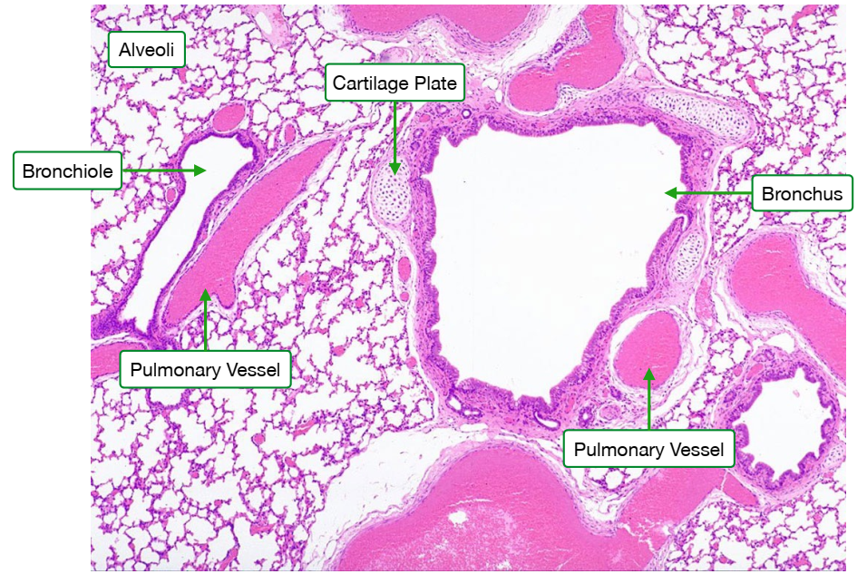
Airway Pathway
- Air travels from the throat to the lungs through a hierarchical branching system of airways:

Main Pathway:
Trachea → Bronchi → Bronchioles → Alveolar Ducts → Alveoli

Trachea (Windpipe)
- Connection: Links the larynx (voice box) to the bronchi.
- Structure:
- Shape: Tube-like.
- Support: C-shaped rings of cartilage maintain an open airway.
- Function:
- Facilitates the movement of air in and out of the lungs.
- Maintains low air resistance during breathing.


Bronchi (Singular: Bronchus)
- Branches: Two primary bronchi branch from the trachea, each entering one lung.
- Structure:
- Support: Irregular blocks of cartilage prevent collapse and keep airways open.
- Function:
- Directs air into each lung.
- Further subdivides to form the bronchial tree, increasing the surface area for air distribution.

Bronchioles
- Description: Smaller branches stemming from the bronchi within each lung.
- Types:
- Terminal Bronchioles:
- Role: Do not participate in gas exchange.
- Function: Supply air to respiratory bronchioles.
- Respiratory Bronchioles:
- Role: Lead to alveolar ducts.
- Function: Supply air directly to alveoli for gas exchange.
- Terminal Bronchioles:
- Structure:
- Lack Cartilage: Unlike larger airways.
- Presence of Smooth Muscle: Controls airway diameter to regulate airflow.


2. Structural Components and Their Functions
Cartilage
- Locations:
- Trachea: C-shaped rings.
- Bronchi: Irregular blocks.
- Functions:
- Airway Support: Keeps airways open, preventing collapse or obstruction during breathing.
- Airflow Efficiency: Reduces air resistance, ensuring smooth passage of air.

Smooth Muscle
- Location: Predominantly in bronchioles.
- Appearance: Thin bands or layers observable under a microscope.
- Functions:
- Airway Regulation: Contracts or relaxes to adjust bronchiole diameter.
- Flow Control: Regulates airflow distribution to different lung regions, optimizing gas exchange.


3. Role and Structure of Alveoli
Alveoli
- Description: Tiny, balloon-like air sacs at the end of the respiratory pathway.
- Surface Area: Approximately 70–75 m² in total, providing a vast area for gas exchange.
- Function:
- Oxygen Uptake: Facilitates the diffusion of oxygen into the blood.
- Carbon Dioxide Removal: Allows carbon dioxide to diffuse out of the blood.
- Importance:
- Efficient Gas Exchange: Large number and surface area compensate for oxygen’s low solubility in water, ensuring effective diffusion necessary for cellular respiration.
