Question 2b (i)
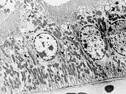
Describe the structure and appearance of the cellular components visible in the electron micrograph. [4]
Answer:

The proximal convoluted tubule (PCT) cells in an electron micrograph typically exhibit the following features:
Basal infoldings: The basal surface of PCT cells often has numerous infoldings. These increase surface area for reabsorption and transport channels and house more mitochondria to provide energy for active transport.
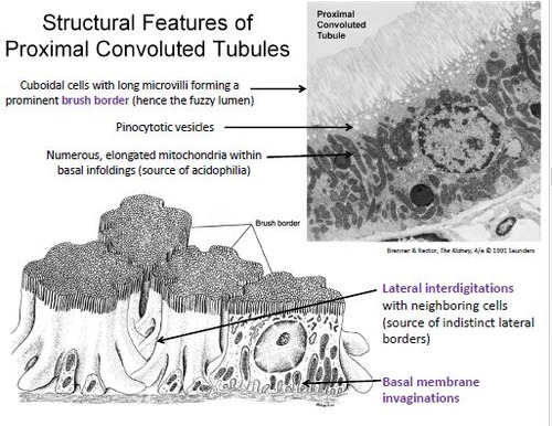
Microvilli: The apical surface of PCT cells contains a dense brush border of microvilli, which appear as a fuzzy or textured layer in the micrograph. These increase surface area, aiding in efficient reabsorption of substances from the filtrate.
Mitochondria: The cells contain numerous mitochondria, which are visible as small, rod-like structures. They provide the necessary ATP for active transport processes in the reabsorption of ions and nutrients.
Tight junctions: Between the PCT cells, tight junctions may be visible. These help maintain a seal between cells, preventing filtrate from leaking between them, ensuring selective reabsorption.