15.07 Muscle Structure and Contraction
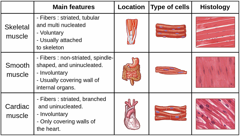
Types of Muscle Tissue
1.Striated Muscle (Skeletal Muscle)
- Location: Attached to skeleton; responsible for voluntary movement.
- Appearance: Striated (striped) under a microscope due to regular arrangement of filaments.
- Control: Neurogenic—requires motor neurone impulses to contract.
2.Cardiac Muscle
- Location: Found exclusively in the heart.
- Appearance: Striated, with cells connected by intercalated discs that facilitate synchronized contraction.
- Control: Myogenic—contracts automatically without neural input.
3.Smooth Muscle
- Location: Found in walls of tubular organs (e.g., blood vessels, digestive tract).
- Appearance: Non-striated (smooth).
- Control: Generally neurogenic but can contract in response to stretch, as seen in arteries.
Structure of Striated Muscle
- Muscle Composition: Each muscle (e.g., biceps) consists of thousands of muscle fibres bundled together and connected to bones by tendons.
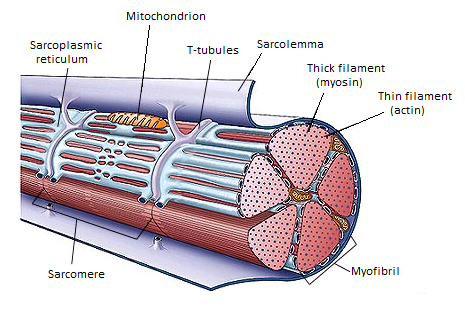
- Muscle Fibre Structure:
- Syncytium: Muscle fibres are multinucleated, formed by fusion of cells.
- Sarcolemma: Cell membrane surrounding each muscle fibre.
- Sarcoplasm: Cytoplasm within the muscle fibre, containing mitochondria and myofibrils.
- Sarcoplasmic Reticulum (SR): Specialized endoplasmic reticulum that stores and releases calcium ions, crucial for contraction.
- T-tubules: Infoldings of the sarcolemma that conduct action potentials into the fibre, ensuring coordinated contraction.
- Myofibrils:
- Composition: Each fibre contains many cylindrical myofibrils made up of thick (myosin) and thin (actin) filaments.
- Striations: Alternating dark (A bands) and light (I bands) regions due to the arrangement of myosin and actin filaments.
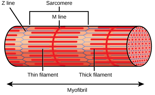
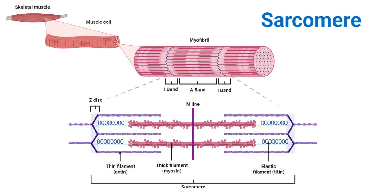
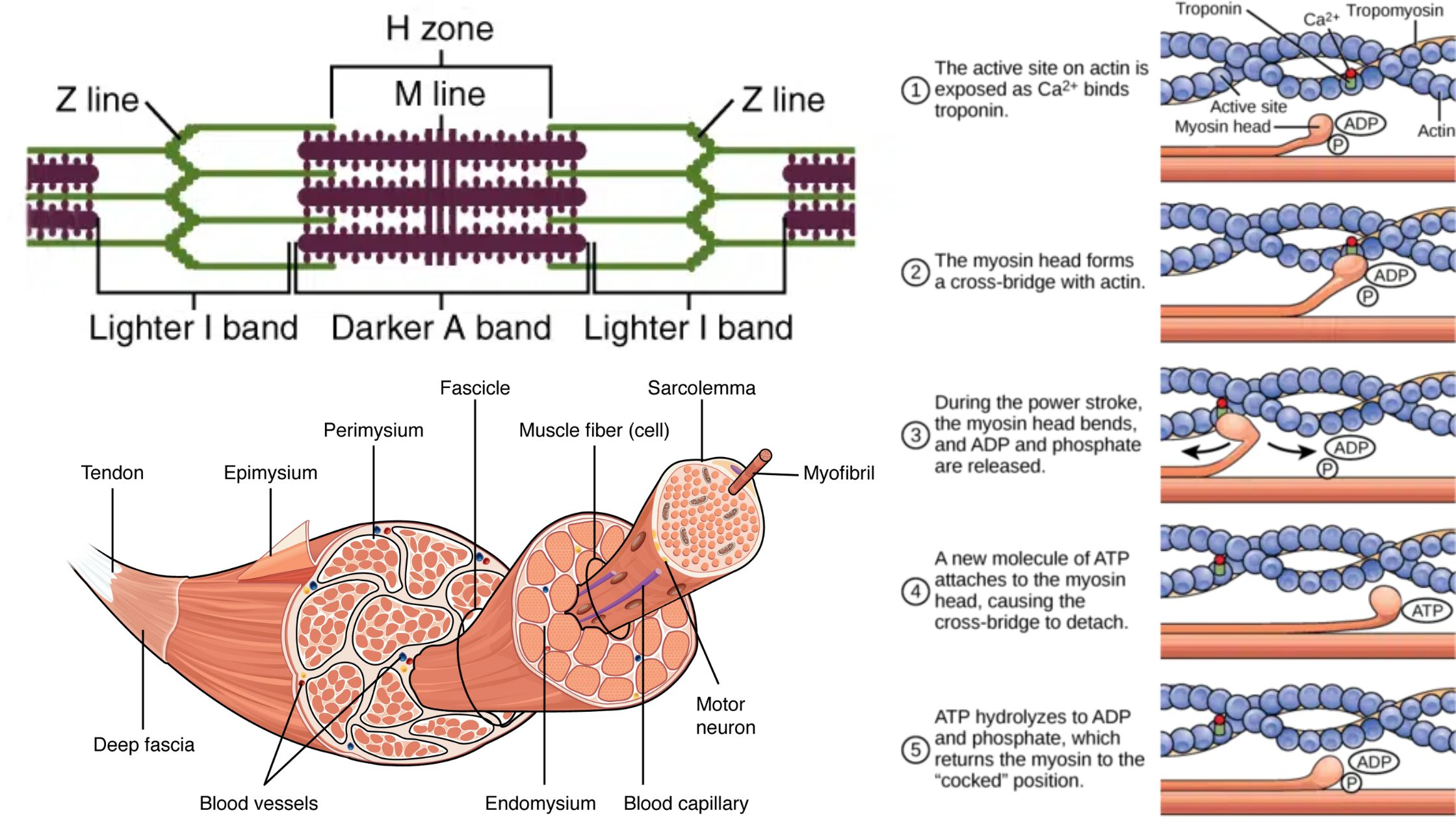
Detailed Myofibril Structure
- Sarcomere: The basic contractile unit of a myofibril, spanning from one Z line to the next.
- Z Line: Anchors actin filaments and marks the boundary of each sarcomere.
- A Band: Dark band containing overlapping actin and myosin filaments.
- I Band: Light band with only actin filaments, no overlap with myosin.
- H Band: Lighter central area within the A band, containing only myosin filaments.
- M Line: Central line within the H band, where myosin filaments are anchored.
Structure and Function of Muscle Filaments
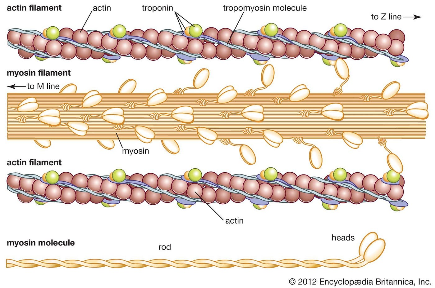
- Thick Filaments (Myosin):
- Composition: Made up of myosin molecules, each with a fibrous tail and a globular head.
- Arrangement: Myosin molecules are bundled with heads pointing away from the M line.
- Myosin Heads: Function as ATPases, breaking down ATP to drive contraction.
- Thin Filaments (Actin):
- Composition: Primarily actin, with regulatory proteins tropomyosin and troponin.
- Actin: Globular proteins linked in a double helix structure, providing binding sites for myosin.
- Tropomyosin: Twists around actin, blocking myosin binding sites in a relaxed muscle.
- Troponin: Calcium-binding protein that, when bound to Ca²⁺, allows tropomyosin to move and expose myosin-binding sites on actin.
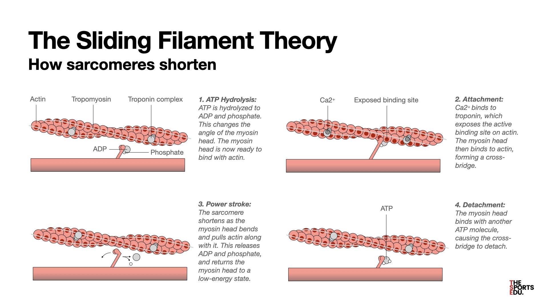
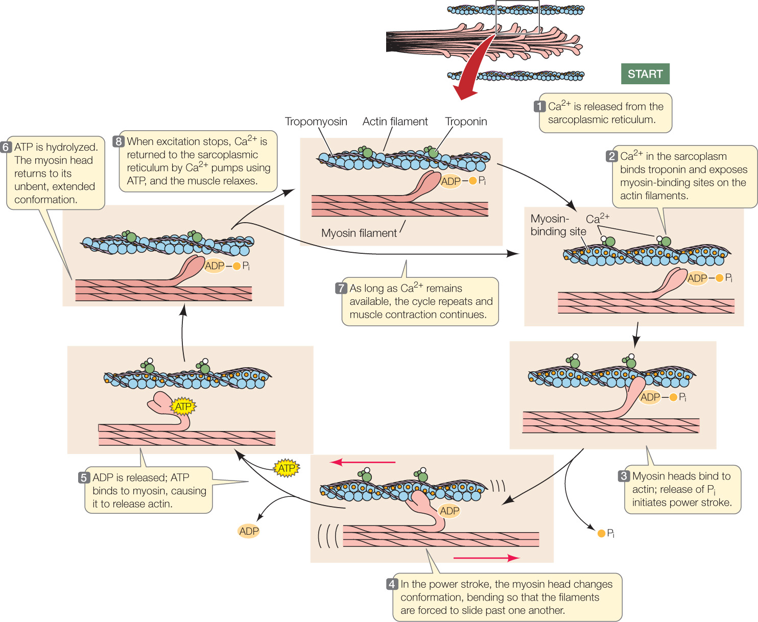
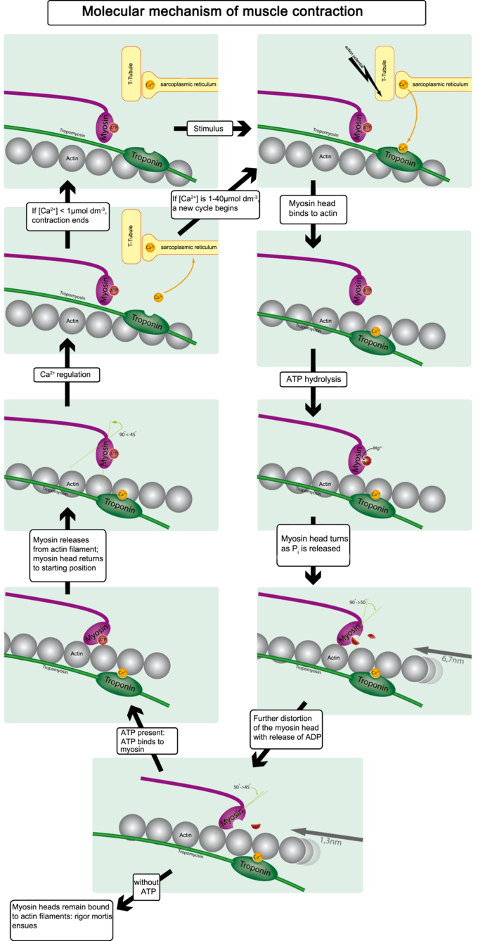
Muscle Contraction: The Sliding Filament Model
1.Muscle Relaxation:
- Tropomyosin and troponin block myosin-binding sites on actin, preventing myosin from attaching.
2.Stimulation and Calcium Release:
- An action potential reaches the neuromuscular junction, where acetylcholine (ACh) is released, causing depolarization of the sarcolemma.
- The action potential travels along T-tubules, triggering Ca²⁺ channels in the SR to open and release calcium ions into the sarcoplasm.
3.Calcium Binding and Cross-Bridge Formation:
- Calcium Ions: Bind to troponin, changing its shape and moving tropomyosin away from myosin-binding sites on actin.
- Cross-Bridge Formation: Myosin heads attach to exposed binding sites on actin.
4.Power Stroke:
- Myosin heads pivot, pulling actin filaments toward the M line and shortening the sarcomere by approximately 10 nm.
5.ATP and Cross-Bridge Cycling:
- ATP Binding: Causes myosin heads to detach from actin.
- ATP Hydrolysis: Resets myosin heads to their original position, ready for the next contraction cycle.
- Continuous Cycle: Repeats as long as Ca²⁺ and ATP are available, further shortening the sarcomere.
6.Muscle Relaxation:
- When stimulation stops, Ca²⁺ ions are pumped back into the SR, tropomyosin re-covers binding sites, and the muscle relaxes.
Neuromuscular Junction and Muscle Activation
- Function: Transmits signals from motor neurones to muscle fibres to initiate contraction.
- Process:
- An action potential reaches the motor end plate, causing an influx of Ca²⁺.
- ACh is released from synaptic vesicles and binds to receptors on the sarcolemma.
- ACh binding opens Na⁺ channels, causing depolarization and triggering an action potential in the muscle fibre.
- This action potential spreads along T-tubules to the SR, resulting in Ca²⁺ release and initiating contraction.
- Termination: Acetylcholinesterase breaks down ACh to prevent continuous stimulation, allowing the muscle to relax when impulses cease.
Energy for Muscle Contraction
- ATP Sources:
- Aerobic Respiration: Provides ATP via mitochondria during sustained, moderate activity.
- Lactate Fermentation: During intense activity, ATP is produced anaerobically, leading to lactate accumulation as a byproduct.
Key Terms
- Sliding Filament Model: Mechanism of muscle contraction where myosin heads pull actin filaments toward the M line, shortening the sarcomere.
- Striated Muscle: Muscle with a striped appearance, due to the arrangement of myofibrils.
- Sarcolemma: Muscle fibre membrane.
- Sarcoplasm: Cytoplasm of muscle fibres, containing mitochondria and myofibrils.
- Sarcoplasmic Reticulum (SR): Stores and releases Ca²⁺ ions essential for muscle contraction.
- Myofibril: Cylindrical bundles of actin and myosin filaments within a muscle fibre.
- Myosin: Thick filament protein, with heads that break down ATP to power contraction.
- Actin: Thin filament protein that interacts with myosin for contraction.
- Sarcomere: Basic contractile unit of a myofibril, between two Z lines.
- Z Line: Anchors actin filaments and separates sarcomeres.
- A Band: Dark band with overlapping actin and myosin.
- I Band: Light band containing only actin.
- H Band: Central part of the A band with only myosin.
- T-tubule: Infolding of sarcolemma that conducts impulses into the muscle fibre.
- Troponin and Tropomyosin: Regulatory proteins controlling actin-myosin binding.
- Neuromuscular Junction: Synapse between a motor neurone and muscle fibre.
Practise Questions
Test 1
1. Which type of muscle tissue is responsible for voluntary movements and is attached to the skeleton?
2. What distinguishes cardiac muscle from skeletal muscle in terms of control mechanisms?
3. Which muscle type is found exclusively in the heart and features intercalated discs?
4. What is the primary role of the sarcoplasmic reticulum (SR) in muscle fibres?
5. In the Sliding Filament Model, what is the basic contractile unit of a myofibril?
6. Which proteins block myosin-binding sites on actin filaments in a relaxed muscle?
7. What initiates the release of calcium ions from the sarcoplasmic reticulum during muscle contraction?
8. During muscle relaxation, what happens to calcium ions?
9. What is the function of intercalated discs in cardiac muscle?
10. Which process terminates the signal at the neuromuscular junction to allow muscle relaxation?
Correct Answers: 0%
Test 2
1. Which type of muscle tissue is found exclusively in the heart and features intercalated discs?
2. What distinguishes smooth muscle from striated and cardiac muscle in terms of appearance?
3. Which component of the muscle fibre is responsible for storing and releasing calcium ions during muscle contraction?
4. In the sliding filament model, what is the role of myosin heads?
5. What is the basic contractile unit of a myofibril?
6. How do troponin and tropomyosin regulate muscle contraction?
7. What initiates the release of calcium ions from the sarcoplasmic reticulum during muscle contraction?
8. During muscle relaxation, what happens to calcium ions?
9. What is the function of intercalated discs in cardiac muscle?
10. Which process terminates the signal at the neuromuscular junction to allow muscle relaxation?
Correct Answers: 0%