15.06 Synapses
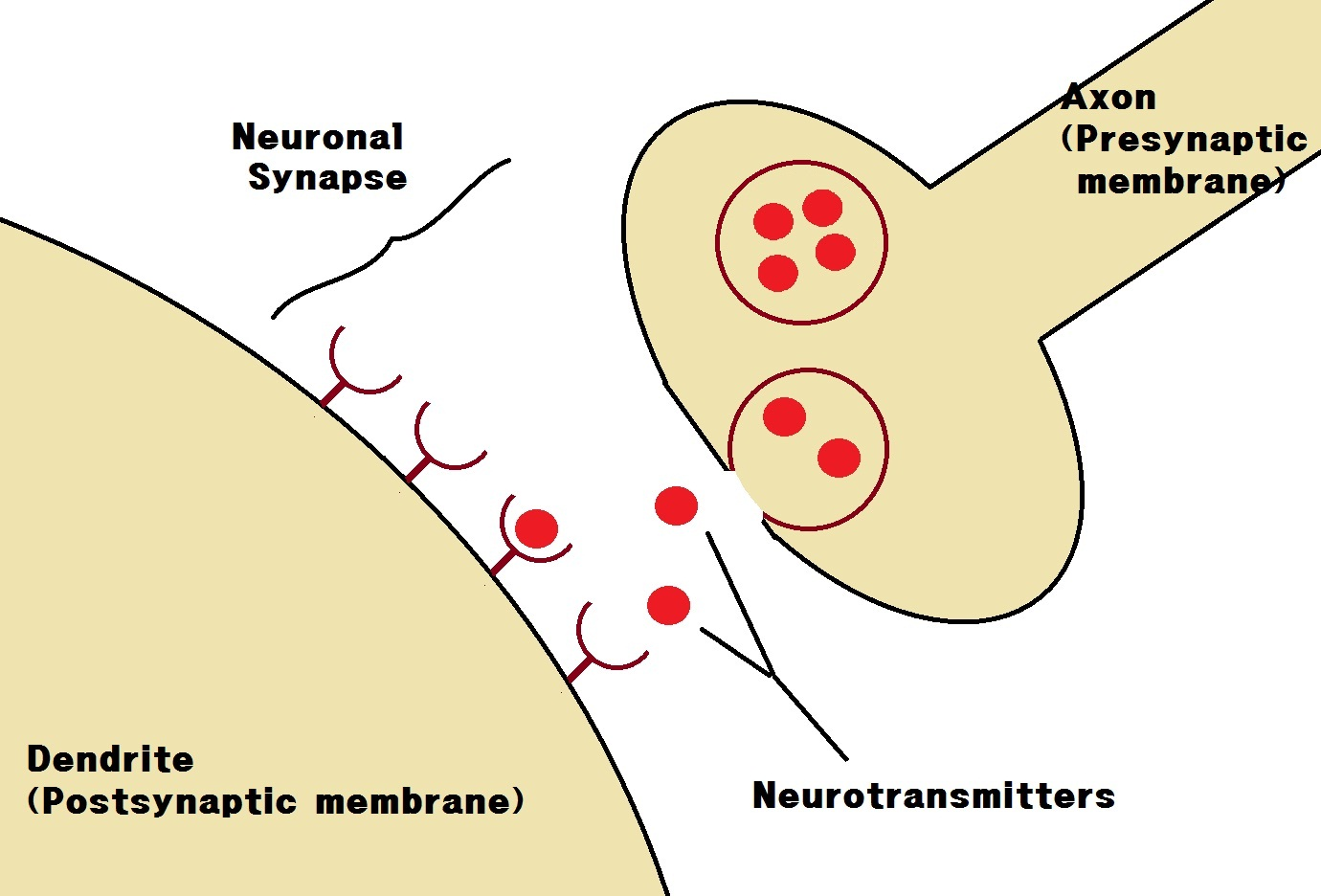
Structure of a Synapse
- Synaptic Cleft: A small 20 nm gap between two neurones where signal transmission occurs.
- Synapse Components:
- Presynaptic Neurone: Neurone ending at the synapse, containing vesicles with neurotransmitter.
- Postsynaptic Neurone: Neurone on the opposite side, with receptor proteins on its membrane.
- Neurotransmitter: Chemical substance that transmits impulses across the synaptic cleft (e.g., acetylcholine).
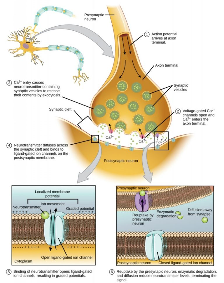
Mechanism of Synaptic Transmission
1.Arrival of Action Potential:
- An action potential reaches the presynaptic neurone’s terminal, depolarizing the membrane.
2.Calcium Ion Influx:
- Depolarization opens voltage-gated calcium channels, allowing Ca²⁺ ions to enter the presynaptic neurone due to a steep electrochemical gradient.
3.Vesicle Fusion and Neurotransmitter Release:
- Ca²⁺ ions cause vesicles containing acetylcholine (ACh) to fuse with the presynaptic membrane, releasing ACh into the synaptic cleft via exocytosis.
4.Diffusion Across Cleft:
- ACh molecules diffuse rapidly (within 0.5 ms) across the cleft to the postsynaptic membrane.
5.Receptor Binding and Depolarization:
- ACh binds to ligand-gated ion channels on the postsynaptic membrane, causing these channels to open and allowing Na⁺ to enter.
- Na⁺ Influx: Depolarizes the postsynaptic membrane; if threshold potential is reached, it triggers an action potential.
6.Enzyme Breakdown of ACh:
- Acetylcholinesterase in the synaptic cleft breaks down ACh into acetate and choline to prevent continuous stimulation.
7.Recycling:
- Choline is reabsorbed by the presynaptic neurone, where it recombines with acetyl coenzyme A to form ACh, ready for the next impulse.
Types of Synapses and Examples
- Cholinergic Synapses: Use ACh as the neurotransmitter, found in the central and peripheral nervous systems.
- Neuromuscular Junction: Specialized synapse between a motor neurone and muscle fibre, leading to muscle contraction.


Functions and Advantages of Synapses
- One-Way Transmission: Ensures impulses travel in one direction only, as neurotransmitter release occurs only from the presynaptic side.
- Signal Integration and Pathway Interconnection:
- Divergence: One neurone can stimulate multiple postsynaptic neurones, allowing one signal to affect multiple effectors (e.g., in reflex responses).
- Convergence: Multiple neurones can stimulate a single postsynaptic neurone, allowing integration of information from various sources, crucial for complex processing in the brain.
- Modulation of Impulse Strength:
- Synapses allow the brain to prioritize and filter signals, enhancing decision-making and coordinated responses.

Synaptic Fatigue
- Synaptic Fatigue: If action potentials arrive continuously, synapses can temporarily deplete their neurotransmitter supply, leading to a temporary inability to transmit signals.
Summary Diagram of Synaptic Transmission

- Acetylcholinesterase breaks down ACh, recycling components back to the presynaptic neurone.
- Action potential arrives at the presynaptic terminal.
- Calcium channels open, allowing Ca²⁺ to enter.
- Synaptic vesicles release neurotransmitter (ACh) by exocytosis.
- ACh diffuses across the synaptic cleft.
- ACh binds to receptors on the postsynaptic membrane, opening Na⁺ channels.
- Depolarization occurs, possibly triggering an action potential.
Key Terms
- Synapse: Junction between two neurones, including the presynaptic and postsynaptic membranes and synaptic cleft.
- Synaptic Cleft: Gap between two neurones where neurotransmitter is released and diffuses across.
- Neurotransmitter: Chemical that transmits nerve impulses across synapses (e.g., acetylcholine, noradrenaline).
- Presynaptic Neurone: The neurone releasing neurotransmitter into the synapse.
- Postsynaptic Neurone: The neurone receiving neurotransmitter, triggering depolarization.
- Acetylcholinesterase: Enzyme that breaks down acetylcholine in the synaptic cleft.
- Neuromuscular Junction: Synapse between a motor neurone and muscle, leading to muscle contraction.
- Ligand-Gated Channel: Protein channel on the postsynaptic membrane that opens in response to neurotransmitter binding.
Practise Questions
Test 1
1. What is the synaptic cleft?
2. Which component of the synapse contains vesicles filled with neurotransmitters?
3. Which neurotransmitter is commonly associated with cholinergic synapses?
4. What initiates the release of neurotransmitters into the synaptic cleft?
5. What is the primary function of acetylcholinesterase in the synaptic cleft?
6. What happens when acetylcholine binds to ligand-gated ion channels on the postsynaptic membrane?
7. What ensures that impulses travel in only one direction across the synapse?
8. What is synaptic fatigue?
9. What is the primary function of the neuromuscular junction?
10. What process terminates the signal at the neuromuscular junction to allow muscle relaxation?
Correct Answers: 0%
Test 2
1. What is the synaptic cleft?
2. Which component of the synapse contains vesicles filled with neurotransmitters?
3. Which neurotransmitter is commonly associated with cholinergic synapses?
4. What initiates the release of neurotransmitters into the synaptic cleft?
5. What is the primary function of acetylcholinesterase in the synaptic cleft?
6. What happens when acetylcholine binds to ligand-gated ion channels on the postsynaptic membrane?
7. What ensures that impulses travel in only one direction across the synapse?
8. What is synaptic fatigue?
9. What is the primary function of the neuromuscular junction?
10. What process terminates the signal at the neuromuscular junction to allow muscle relaxation?
Correct Answers: 0%