15.11 End of Chapter Questions
Question 1
ADH, insulin, and glucagon are key hormones involved in maintaining homeostasis. ADH is a short peptide, glucagon is a single polypeptide, and insulin consists of two polypeptides. The target cells for these hormones only respond if they possess specific receptors on their cell surfaces.
a.i) Explain why it is essential for the target cells of ADH, insulin, and glucagon to have specific receptors on their surfaces to be able to respond to these hormones. [2]
a) Why target cells for ADH, insulin, and glucagon need specific cell surface receptors to respond:
Signal Transduction: Once the hormone binds to its receptor, it triggers a series of intracellular signals, often through a secondary messenger system (e.g., cAMP for glucagon and insulin), that cause the target cell to respond appropriately. Without these receptors, the target cells would not be able to detect or respond to the hormone.
Hormone Action Mechanism: Hormones, including ADH, insulin, and glucagon, act as chemical messengers that regulate various physiological processes. For a hormone to have an effect on a target cell, it must bind to a specific receptor on the surface or inside of the target cell.
Receptors Are Highly Specific: The receptors on the target cells are protein molecules with a specific shape that can only bind to their complementary hormone. This “lock-and-key” mechanism ensures that only the correct hormone will interact with the correct receptor.
a.ii) Identify the target cells for each of these hormones. [3]
b) Target Cells for Each Hormone:
ADH (Antidiuretic Hormone):
- Target Cells: ADH mainly affects cells in the kidneys, particularly in the distal convoluted tubules and collecting ducts.
- These cells have receptors for ADH, which allow for the regulation of water reabsorption, influencing the concentration of urine and body hydration.
Insulin:
- Target Cells: Insulin primarily targets muscle cells, fat cells (adipocytes), and liver cells (hepatocytes).
- These cells have insulin receptors that allow them to take in glucose from the blood, reducing blood glucose levels.
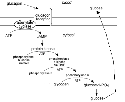
Glucagon:
- Target Cells: Glucagon mainly targets liver cells (hepatocytes), where it promotes the breakdown of glycogen into glucose (glycogenolysis) and the production of glucose from non-carbohydrate sources (gluconeogenesis), thus increasing blood glucose levels.
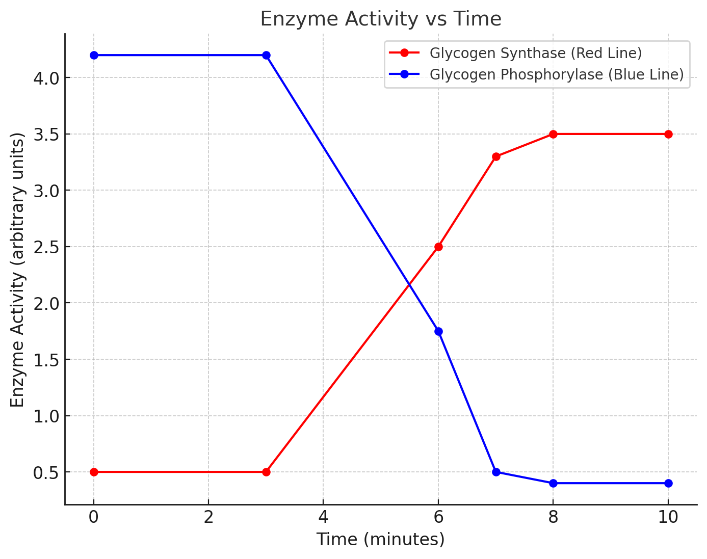
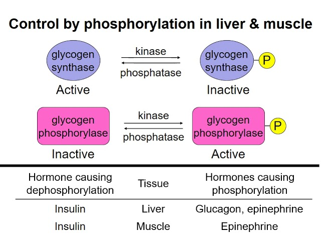
Two enzymes involved in the synthesis and breakdown of glycogen in liver cells are glycogen synthase and glycogen phosphorylase. A solution with a high glucose concentration was used to expose liver cells, and the activity of both enzymes was measured over a period of 10 minutes. The following graph shows the results.
a) Describe the changes in enzyme activity for both glycogen synthase and glycogen phosphorylase as shown in the graph. [2]
- Glycogen Phosphorylase (Blue Line):
- The activity of glycogen phosphorylase starts at 4.2 arbitrary units and remains constant until 3 minutes.
- From 3 to 6 minutes, its activity decreases almost linearly, dropping to 1.75 arbitrary units.
- Between 6 and 7 minutes, the decline continues, reaching 0.5 arbitrary units.
- Finally, between 7 and 8 minutes, the enzyme activity drops sharply to 0.4 arbitrary units, where it stays constant between 8 and 10 minutes.
- Glycogen Synthase (Red Line):
- The activity of glycogen synthase begins at 0.5 arbitrary units and remains steady until 3 minutes.
- After 3 minutes, its activity increases gradually, reaching 2.5 arbitrary units between 3 and 6 minutes.
- The rate of increase slows between 6 and 7 minutes, peaking at 3.3 arbitrary units.
- Between 8 and 10 minutes, the enzyme activity stabilizes at 3.5 arbitrary units.
b) What happens to the amount of glycogen stored in a liver cell between 4 minutes and 10 minutes? [1]
- Glycogen storage increases between 4 and 10 minutes because glycogen synthase activity is increasing while glycogen phosphorylase activity is decreasing, which favours the synthesis of glycogen and reduces its breakdown.
c) After a meal rich in starch, similar changes in the activity of glycogen synthase and glycogen phosphorylase are observed in liver cells.
Explain why the activities of these two enzymes change following such a meal. [4]
- After consuming a starch-rich meal, blood glucose levels rise.
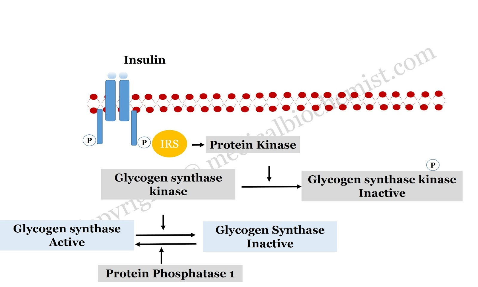
- The increased glucose concentration stimulates the pancreas to release insulin, which promotes glycogen synthesis by activating glycogen synthase.
- At the same time, insulin inhibits the activity of glycogen phosphorylase, an enzyme responsible for breaking down glycogen into glucose, ensuring that glucose is stored rather than released into the bloodstream.
- This coordinated action helps lower blood glucose levels by promoting storage in the liver.
d) Cells can regulate the activity of enzymes.
Describe how the activity of glycogen phosphorylase is controlled. [2]
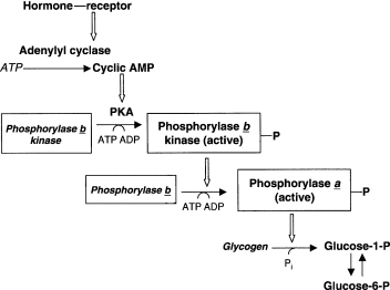
- The activity of glycogen phosphorylase is regulated by phosphorylation.
- When glycogen phosphorylase is phosphorylated (by enzymes like phosphorylase kinase), it becomes active and promotes glycogen breakdown.
- In contrast, when glycogen phosphorylase is dephosphorylated (by the enzyme phosphatase), it becomes inactive, stopping the breakdown of glycogen.
- This regulation ensures that glycogen is only broken down when needed, such as during low blood glucose levels.
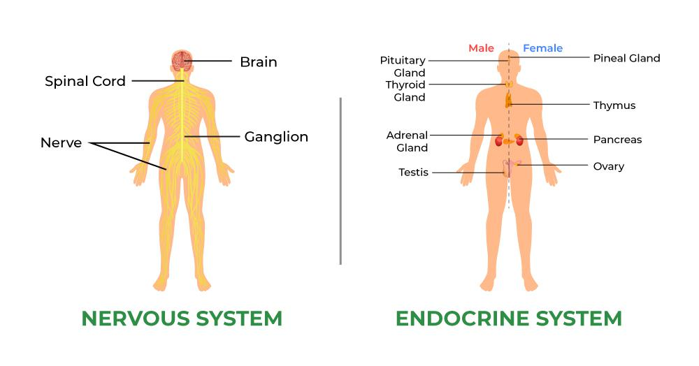
e) Why is the endocrine system more suitable than the nervous system for controlling the effectors that regulate blood composition? [3]
- The endocrine system is ideal for controlling blood composition because it uses hormones that are released into the bloodstream and can act on multiple target organs over a longer period.
- This allows for long-term regulation, such as maintaining glucose levels or water balance.
- In contrast, the nervous system is faster but is better suited for short-term, rapid responses like reflexes or immediate actions.
- The hormonal control provided by the endocrine system ensures sustained, widespread regulation of internal conditions, such as blood glucose, which the nervous system cannot coordinate as effectively over longer periods.
Question 2
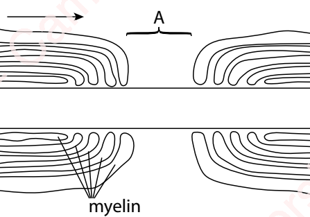
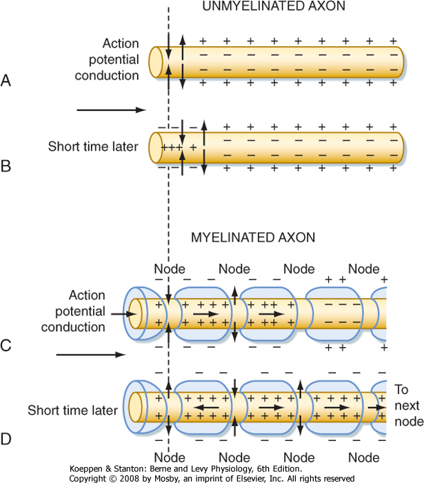
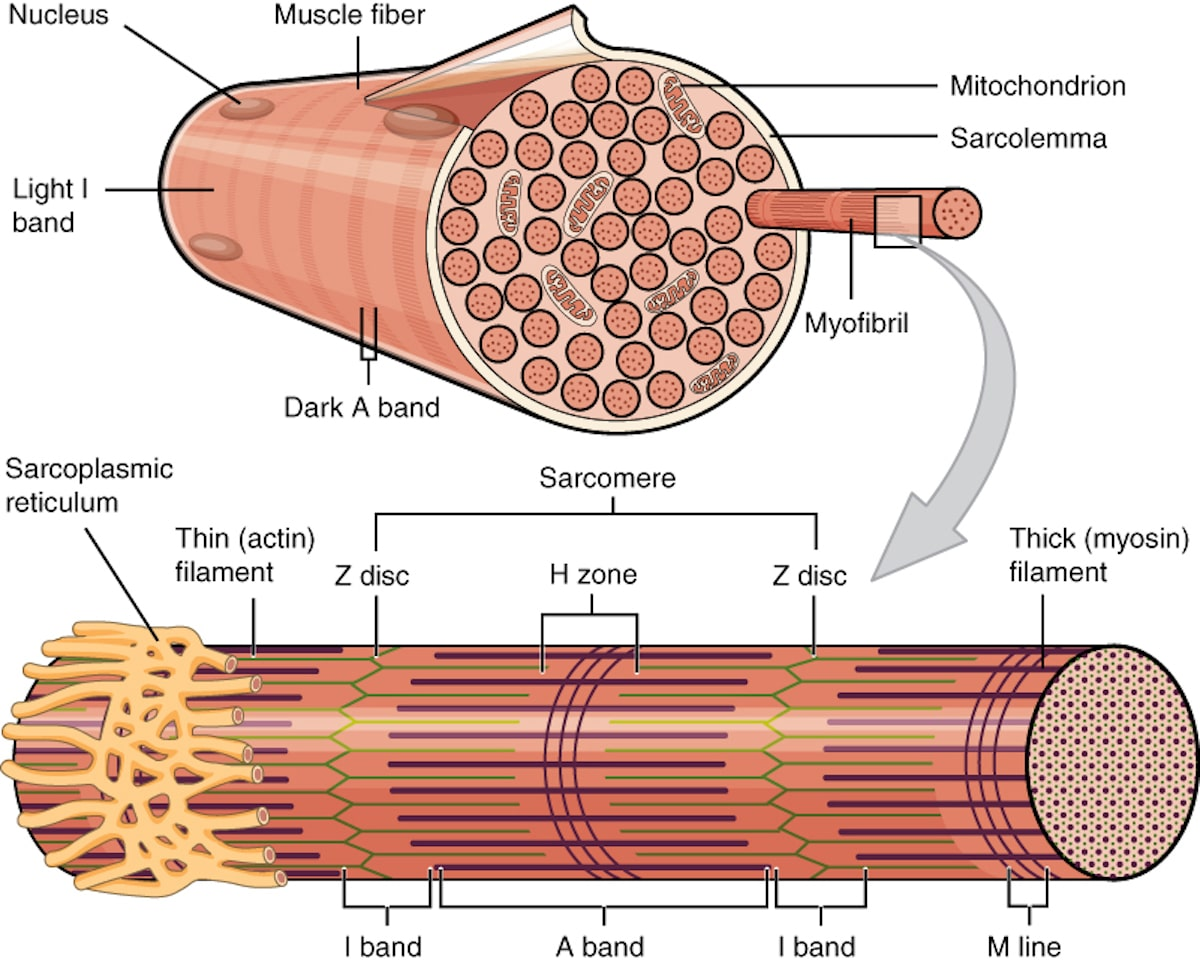
The following diagram is taken from a transmission electron microscope (TEM) image of a longitudinal section through a myelinated neuron. The diagram shows the region labeled A, which involves myelin sheaths around an axon.
a) i) What is the name of the region of the neuron labeled A? [1]
ii) What is the name of the cell responsible for producing myelin? [1]
i) The region labeled A is the Node of Ranvier on the axon.
ii) The cell responsible for producing myelin is the Schwann cell.
b) How does myelin contribute to the transmission of nerve impulses? [4]
- Myelin acts as an insulator around the axon, preventing ion flow across the membrane in the myelinated areas.
- This insulation increases the speed of impulse transmission by allowing the action potential to jump from one node of Ranvier to the next in a process called saltatory conduction.
- The myelin sheath also reduces energy consumption during impulse transmission, as fewer ions need to be actively transported back into the neuron after depolarization.
- Overall, the presence of myelin makes nerve signal transmission much faster and more efficient compared to unmyelinated neurons.
c) Describe the changes that occur at region A during the passage of an impulse in the direction indicated by the arrow. [5]
- The myelin sheath prevents ion movement in the insulated regions, allowing the action potential to “jump” from node to node in a process known as saltatory conduction.
- At the node of Ranvier (region A), the action potential is regenerated at each node, ensuring rapid propagation of the impulse.
- When an impulse reaches the node of Ranvier (the gap between two Schwann cells), the axon membrane depolarizes as sodium ions (Na⁺) rush in through voltage-gated channels.
- This depolarization creates a local electrical current, which triggers the next set of sodium channels downstream to open, continuing the depolarization.
- As the action potential propagates along the axon, potassium ions (K⁺) move out of the axon, restoring the resting potential behind the action potential.
Question 3
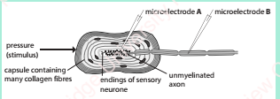
The Pacinian corpuscle is a type of receptor found in the skin’s dermis. It contains the sensory neuron’s ending, which is encased by several layers of connective tissue called a capsule. The activity of the Pacinian corpuscle was investigated by inserting microelectrodes into the axon at the positions shown in the diagram.
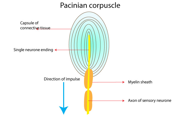
a) What changes likely occurred in the unmyelinated part of the axon when pressure was applied to the Pacinian corpuscle? [4]
- As pressure was applied to the Pacinian corpuscle, the mechanical stimulus deformed the capsule, which in turn activated the sensory nerve endings.
- In the unmyelinated region of the axon, the deformation would likely cause a depolarization of the sensory neuron’s membrane, triggering action potentials.
- This local depolarization would propagate along the axon as a wave of depolarization, eventually reaching the myelinated region.
- The action potentials generated in this region would travel through the unmyelinated part to be transmitted further, signaling the presence of pressure.
b) How can the pattern of recordings from microelectrode B be explained as pressure on the Pacinian corpuscle increases? [4]
- As pressure on the Pacinian corpuscle increases, it likely causes a greater deformation of the capsule, which leads to a stronger stimulus at the sensory nerve endings.
- This increased stimulus would result in a greater frequency of action potentials being generated at the unmyelinated region of the axon, recorded at microelectrode B.
- Initially, there may be a gradual increase in firing rate as pressure builds up, reflecting a graded response to stimulus intensity.
- Once the pressure reaches a certain level, the action potentials recorded at microelectrode B may reach a saturation point, where the frequency of action potentials stabilizes, indicating maximum response to the applied pressure.
c) Why are the sensory neurones of Pacinian corpuscles myelinated rather than unmyelinated? [3]
- Faster signal transmission is crucial for the rapid response to pressure changes, which is the primary function of the Pacinian corpuscle in detecting mechanical stimuli.
- Myelination in sensory neurons of Pacinian corpuscles ensures that action potentials are transmitted faster, allowing for quick detection of pressure changes.
- The myelin sheath allows for saltatory conduction, where the action potential jumps from one node of Ranvier to the next, speeding up the transmission of nerve signals compared to unmyelinated axons.
Question 4
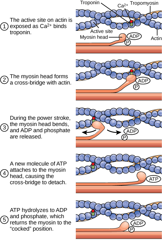
a) Describe the sequence of events that occur when an impulse crosses a synapse between two neurons. [5]
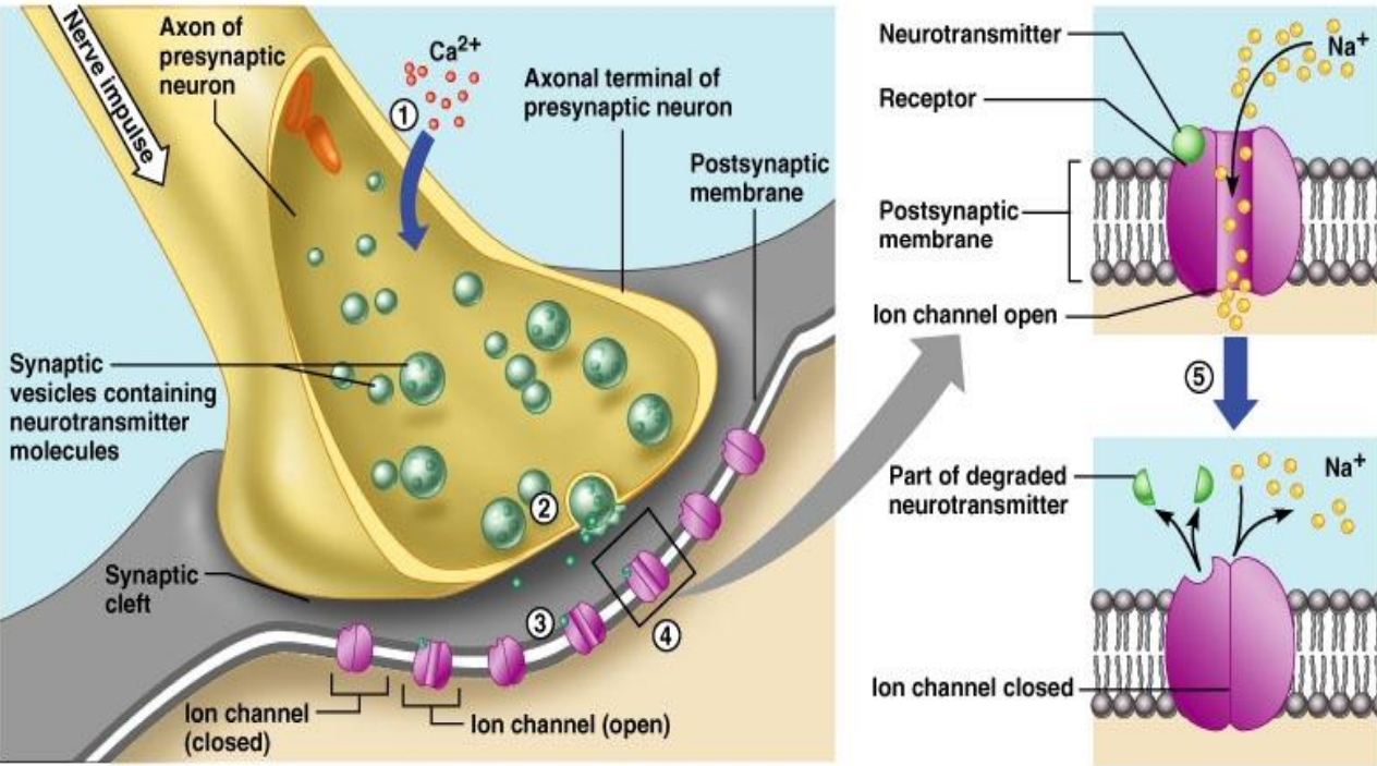
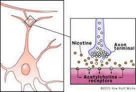
- When an action potential reaches the presynaptic terminal, it causes the calcium ion channels to open.
- Calcium ions (Ca²⁺) flow into the presynaptic neuron, triggering the vesicles containing neurotransmitters (e.g., acetylcholine) to move toward and fuse with the presynaptic membrane.
- The neurotransmitter is released into the synaptic cleft by exocytosis.
- The neurotransmitter binds to receptor proteins on the postsynaptic membrane, causing ligand-gated ion channels to open.
- This leads to a change in the postsynaptic membrane potential, generating a new action potential in the postsynaptic neuron if the threshold is reached.
- The neurotransmitter is broken down by enzymes (e.g., acetylcholinesterase) or taken back up into the presynaptic neuron to terminate the signal.
b) The table below shows the effects of four compounds at cholinergic synapses.
| Compound | Effect at cholinergic synapses |
|---|---|
| Curare | Competes with ACh for its receptor site on ligand-gated sodium ion channel proteins |
| Eserine | Competes with ACh for the active site of acetylcholinesterase |
| Methylmercury | Inhibits the enzyme that synthesizes ACh |
| Nicotine | Activates some ligand-gated sodium ion channels on postsynaptic membranes |
i) For each compound, state and explain its effect on transmission across cholinergic synapses. [8]
Curare:
- Effect: Curare blocks acetylcholine (ACh) from binding to its receptor sites on the postsynaptic membrane.
- Explanation: By competing with ACh for the receptor sites on ligand-gated sodium ion channels, curare prevents the opening of these channels, thus inhibiting synaptic transmission and muscle contraction.
Eserine:
- Effect: Eserine inhibits the enzyme acetylcholinesterase.
- Explanation: Acetylcholinesterase normally breaks down ACh in the synaptic cleft. By inhibiting this enzyme, eserine prolongs the action of ACh at the synapse, enhancing neurotransmission and possibly leading to overstimulation of the postsynaptic neuron.
Methylmercury:
- Effect: Methylmercury inhibits the enzyme that synthesizes ACh.
- Explanation: By blocking the synthesis of ACh, methylmercury reduces the amount of neurotransmitter available for release into the synaptic cleft, impairing synaptic transmission and disrupting normal neural communication.
Nicotine:
- Effect: Nicotine activates some ligand-gated sodium ion channels on postsynaptic membranes.
- Explanation: By binding to and activating nicotinic receptors, nicotine causes the opening of sodium ion channels, leading to depolarization of the postsynaptic membrane, which can mimic the effect of ACh and enhance synaptic transmission.
ii) Explain how eserine can counteract the effect of curare. [3]
- Eserine counters curare’s effect by inhibiting acetylcholinesterase, the enzyme responsible for breaking down acetylcholine (ACh) in the synaptic cleft.
- With acetylcholine remaining in the synapse longer due to the inhibition of acetylcholinesterase, it can outcompete curare for binding to the receptor sites on the postsynaptic membrane.
- As a result, the ACh can still activate the sodium ion channels despite the presence of curare, partially reversing the blocking effect of curare and restoring normal transmission.
Question 5
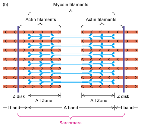
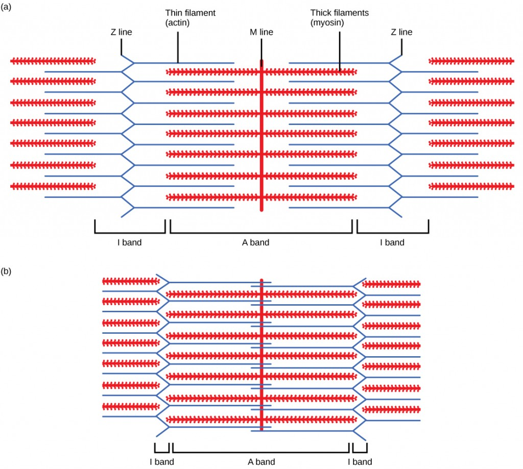
a) i) Identify the parts labeled K, L, and M in the electron micrograph of the myofibrils in a relaxed muscle. [3]
ii) How many myofibrils can be seen in the electron micrograph? Justify your answer. [2]
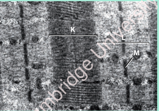
i) The parts labeled K, L, and M are:
- L: The Z-line (also known as the Z-disc), where the actin filaments of adjacent sarcomeres are anchored.
- K: The A-band, which contains the entire length of the thick (myosin) filaments.
- M: The I-band, which is the region between two A-bands containing only thin (actin) filaments.
ii) The number of myofibrils visible in the electron micrograph depends on the specific image, but typically, several myofibrils are visible in a section of muscle.
The number can be determined by counting distinct bundles of thick and thin filaments across the image.
In this case we see 1 myofibril since we are looking at one sarcomere pattern.
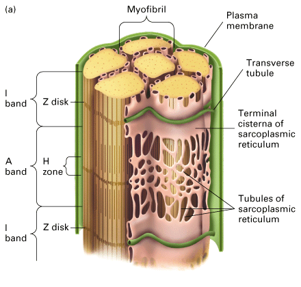
b) i) The electron micrograph shows many glycogen granules and mitochondria. Why are these structures present in muscle cells? [2]
- Glycogen granules are present in muscle cells as a form of energy storage.
- During periods of muscle activity, glycogen is broken down into glucose, which is used to produce ATP for muscle contraction.
- Mitochondria are present because they are the powerhouses of the cell, producing ATP via aerobic respiration, which is needed for sustained muscle activity and contraction.
ii) How can you tell that the electron micrograph shows relaxed muscle rather than contracted muscle? [3]
- The sarcomeres in relaxed muscle appear longer compared to contracted muscle, where the sarcomeres would be shortened due to the sliding filament mechanism.
- The I-bands and H-zones in relaxed muscle are more prominent, as the actin and myosin filaments are not fully overlapped.
- The Z-lines are spaced further apart in relaxed muscle, indicating that the muscle fibers are not contracted.

c) The electron micrograph is magnified 16,000 times. Calculate the actual length of the sarcomere that includes the region labeled K. Provide your answer in micrometers (µm). [2]
To calculate the actual length of the sarcomere, the formula used is:
Actual length = Length on micrograph/Magnification
The actual length of the sarcomere can be obtained by measuring the length of the sarcomere in the image and dividing that by the magnification (16,000). The final result will be in micrometers (µm).
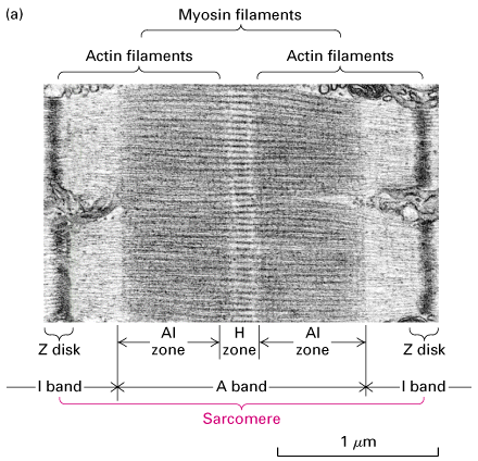
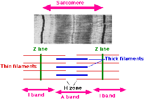
Question 6
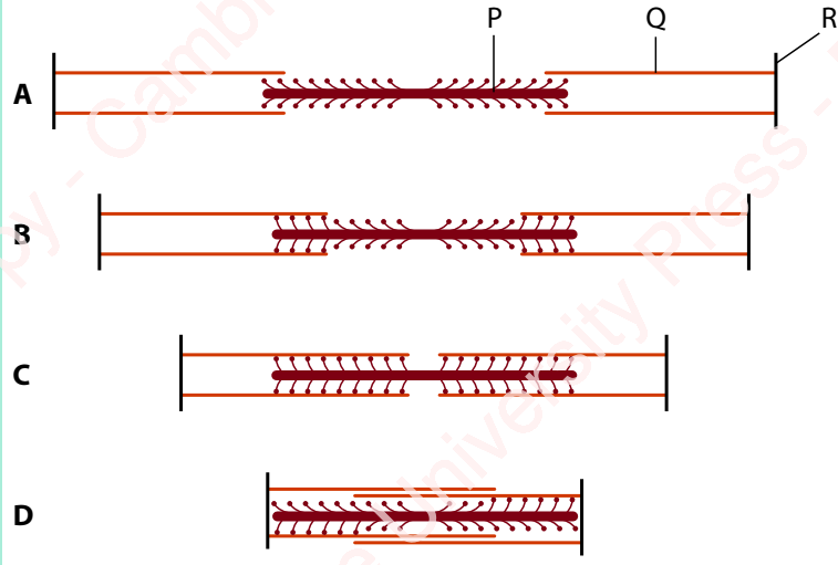
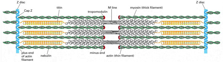
a) Identify the parts labeled P, Q, and R in the sarcomere diagrams. [3]
R: Z-discs (the boundary lines of the sarcomere)
P: Myosin (the thick filament)
Q: Actin (the thin filament)
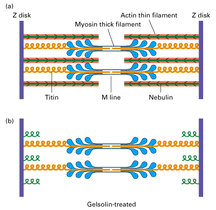
b) Why are no actin–myosin cross-bridges visible in diagram A? [2]
In diagram A, which represents the relaxed state of the sarcomere, the myosin heads are not in contact with the actin filaments because the actin-binding sites on the actin filaments are blocked by tropomyosin. This prevents the formation of cross-bridges between actin and myosin.
c) Muscle fibers can generate more force in some contraction states than others. Which diagram shows the state that can develop the greatest force? Explain why. [4]
In diagrams C and D, the overlap is too short or too long, reducing the number of effective cross-bridges and thus the force produced.
Diagram B shows the state of contraction that can generate the greatest force.
This is because in Diagram B, the actin and myosin filaments overlap at an optimal length, allowing for the maximum number of cross-bridges to form.
When the filaments are at this ideal overlap, the force generated during contraction is maximized.
d) Why would the sarcomere in diagram D be unable to contract any further? [1]
In Diagram D, the sarcomere is at maximum contraction, and the Z-discs are very close to the M-line.
At this point, the actin filaments overlap fully with the myosin filaments, and no further shortening is possible because the actin filaments are already as close as they can get to the M-line.
e) A muscle can contract with force, but it cannot pull itself back to its original relaxed length.
i) With reference to the mechanism of muscle contraction, explain why this is so. [2]
During muscle contraction, ATP is used to power the myosin heads to form cross-bridges with actin and slide the actin filaments towards the M-line.
Elastic components (like titin and the connective tissue) help the muscle return to its resting length, but the muscle itself cannot actively reverse the contraction without additional force.
However, when the muscle relaxes, it requires ATP to actively pump calcium ions back into the sarcoplasmic reticulum, and this process doesn’t provide the energy to fully return the muscle to its original length.
ii) How could the sarcomere in diagram D be returned to the state shown in diagram A? [2]
To return the sarcomere to the relaxed state shown in Diagram A, the calcium ions need to be pumped back into the sarcoplasmic reticulum.
This removes the calcium ions from the actin filaments, causing the tropomyosin to block the actin-binding sites, thus preventing further actin-myosin cross-bridge formation and allowing the sarcomere to return to its relaxed length.
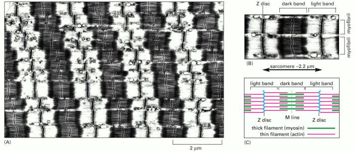
Question 7
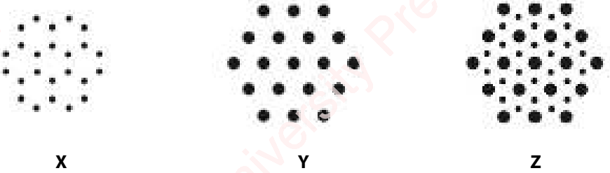
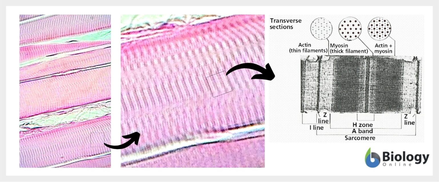
a) Describe the differences between sections X, Y, and Z. You may use a labelled diagram to illustrate your answer. [4]
Section X:
- The structure shows a group of small dots arranged in a hexagonal pattern. This arrangement resembles the triad of myosin filaments, which are associated with actin filaments and appear as small circular patterns in the cross section of a myofibril.
- Diagram X represents a cross-section where the actin filaments are arranged in a flower-like pattern, typical of the thin filaments of the myofibril.
Section Y:
- The section shows a series of larger black dots arranged in a hexagonal shape, with the base of the hexagon having 3 dots, followed by 4, then 5, and back to 4 and 3 dots.
- This arrangement is characteristic of myosin filaments in the A-band of the sarcomere, where thick myosin filaments are present in a regular, repeating structure.
Section Z:
- Section Z is similar to Y but has a hexagonal structure of smaller dots surrounding each of the larger dots.
- This represents the myosin filaments, with each larger dot representing a myosin head and the surrounding smaller dots indicating actin filaments. This cross-section shows the actin-myosin overlap that is crucial for muscle contraction.
b) The sections were taken from a relaxed muscle fibre. Suggest how the sections would appear if taken from a fibre that had contracted to its maximum extent. Explain your answer. [3]
In a contracted muscle fibre, the sarcomere length would decrease.
- Section X would appear more tightly packed, with the actin filaments overlapping more with myosin filaments, reducing the spacing between them.
- Section Y would show a shortened A-band, as the thick myosin filaments are closer together in a contracted state.
- Section Z would show greater overlap between actin and myosin filaments, leading to the Z-lines moving closer to the M-line. The hexagonal arrangement would appear compressed, indicating maximal contraction.
c) Muscle weakness in racehorses may sometimes be related to a deficiency of calcium. Outline the roles of calcium ions in the coordination of muscle contraction. [6]
- Calcium ions bind to troponin: When a muscle is stimulated, calcium ions are released from the sarcoplasmic reticulum. These calcium ions bind to the troponin complex, which is attached to the actin filaments.
- Troponin-tropomyosin complex changes shape: The binding of calcium ions to troponin causes a conformational change in the troponin-tropomyosin complex, which moves tropomyosin away from the actin-binding sites on the actin filament.
- Cross-bridge formation: With the actin-binding sites exposed, the myosin heads can attach to the actin filaments and form cross-bridges. This is the beginning of the sliding filament mechanism.
- ATP hydrolysis and power stroke: The myosin heads use ATP to undergo a power stroke, pulling the actin filaments towards the center of the sarcomere and causing muscle contraction.
- Relaxation of muscle: When the nerve stimulation stops, calcium ions are actively pumped back into the sarcoplasmic reticulum. This removes calcium from troponin, causing the tropomyosin to block the actin-binding sites again, leading to muscle relaxation.
- Muscle weakness from calcium deficiency: If there is a calcium deficiency, the muscle’s ability to contract efficiently is impaired. This is because there is not enough calcium available to bind to troponin, meaning the cross-bridge cycle cannot occur effectively, leading to muscle weakness.
Question 8
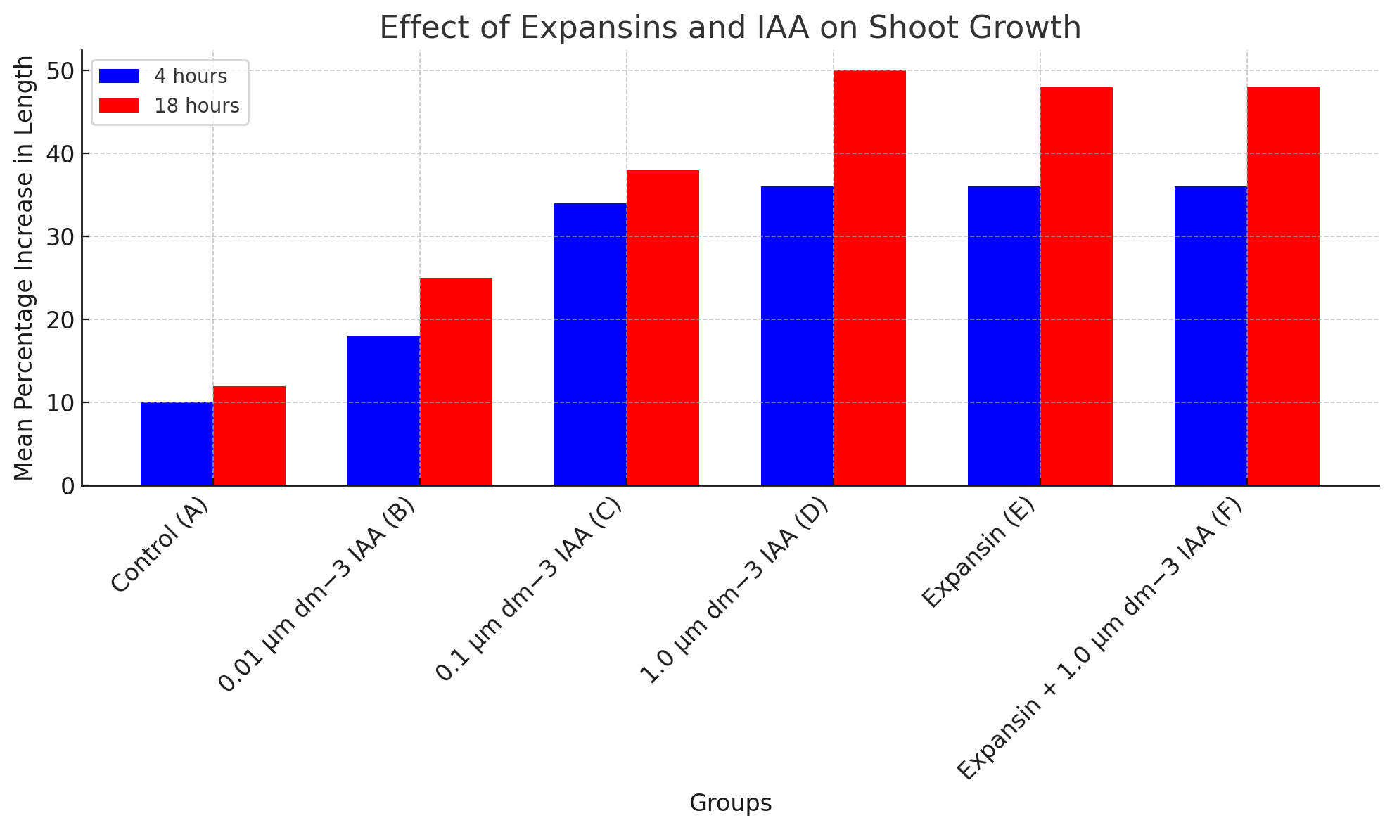
a) The experiment investigated the effects of expansins and auxin (IAA) on shoot growth in Arabidopsis thaliana. Based on the bar chart, describe the results of the experiment at 4 hours and 18 hours. [4]
At 4 hours (blue bars):
- The control group (A) showed a modest increase in length of about 10%.
- The group treated with 0.01 µm dm⁻³ IAA (B) had a slightly higher increase, reaching 18%.
- The group treated with 0.1 µm dm⁻³ IAA (C) showed an increase of about 34%.
- The group treated with 1.0 µm dm⁻³ IAA (D) had a significant increase, reaching 36%.
- The group treated with expansins (E) showed a 36% increase, similar to group D.
- The group treated with expansins + 1.0 µm dm⁻³ IAA (F) showed the same increase as group E, 36%.
At 18 hours (red bars):
- The control group (A) showed a higher increase, reaching 12%.
- The group treated with 0.01 µm dm⁻³ IAA (B) had a 25% increase.
- The group treated with 0.1 µm dm⁻³ IAA (C) reached 38%.
- The group treated with 1.0 µm dm⁻³ IAA (D) reached 50%, showing the highest increase.
- The group treated with expansins (E) had a similar increase as group D, with 48%.
- The group treated with expansins + 1.0 µm dm⁻³ IAA (F) also showed a 48% increase, identical to group E.
b) What do the results suggest about the combined effect of expansins and IAA on shoot growth? [3]
The combination of expansins and IAA in group F resulted in a similar increase in shoot length (48%) as the group treated with expansins alone (E), both at 18 hours.
This suggests that expansins alone may be sufficient to achieve the maximum increase in shoot growth, and the presence of IAA does not further enhance the effect when combined with expansins.
The results imply that the effect of expansins on shoot growth may be independent of IAA at the concentrations tested in this experiment.
c) Based on the bar chart, describe the effect of increasing IAA concentration on shoot growth. [3]
As the concentration of IAA increases, the percentage increase in shoot length also increases, but only up to a certain point.
At 0.01 µm dm⁻³ IAA (group B), there is a modest increase in growth (18% at 4 hours, 25% at 18 hours).
At 0.1 µm dm⁻³ IAA (group C), the growth increases to 34% at 4 hours and 38% at 18 hours.
The highest concentration of 1.0 µm dm⁻³ IAA (group D) results in the greatest growth increase, reaching 36% at 4 hours and 50% at 18 hours.
These results show a positive correlation between IAA concentration and shoot growth, with the greatest effect observed at the highest IAA concentration.
d) What can be concluded about the role of expansins in plant growth based on the results shown in the bar chart? [3]
Expansins have a significant role in promoting shoot growth, as shown by the 36% increase in length at 4 hours and 36% at 18 hours in group E, where expansins alone were applied.
The results suggest that expansins likely play a role in cell wall loosening, which facilitates cell expansion and contributes to growth.
The absence of further growth increase when expansins are combined with high concentrations of IAA (group F) suggests that expansins alone may be sufficient to maximize the growth effect.
Question 9
Investigate the effect of different concentrations of gibberellin on the activity of amylase in germinating barley grains. Outline a strategy for conducting this investigation.
To investigate the effect of different concentrations of gibberellin on the activity of amylase in germinating barley grains, you can follow this strategy:
Preparation of barley grains:
Obtain a uniform batch of barley grains and allow them to germinate under controlled conditions (e.g., temperature, humidity) for a set period to ensure consistent starting conditions.
Preparation of gibberellin solutions:
Prepare a series of gibberellin solutions with different concentrations. For example, prepare concentrations such as 0 (control), 0.1 μmol dm⁻³, 0.5 μmol dm⁻³, 1.0 μmol dm⁻³, 5.0 μmol dm⁻³, and 10.0 μmol dm⁻³ to test a range of gibberellin concentrations.
Application of gibberellin:
For each treatment group, spray or soak the germinating barley grains with the respective gibberellin solution. Ensure that the grains in the control group are treated with water or a gibberellin-free solution to account for any other environmental variables.
Allow the grains to continue germinating:
Allow the barley grains to germinate further for a fixed period (e.g., 24–48 hours) after the gibberellin treatment, ensuring uniform exposure to the hormone across all groups.
Extraction of amylase:
After the germination period, extract amylase from the barley grains by grinding or crushing the grains and then using a suitable extraction buffer to separate the enzyme.
Measuring amylase activity:
Test the amylase activity by measuring the breakdown of starch, which can be done using an iodine test or by quantifying the amount of maltose produced using a colorimetric assay or a glucose oxidase method.
Control variables:
Keep all other variables constant (e.g., temperature, light exposure, germination time) across all groups to ensure that any differences in amylase activity are due to gibberellin concentration.
Data analysis:
Record the results of amylase activity for each gibberellin concentration. Plot a graph of amylase activity (y-axis) against gibberellin concentration (x-axis).
Analyze the relationship to determine whether higher concentrations of gibberellin correlate with increased amylase activity.
Conclusion:
Draw conclusions based on the data, determining the concentration of gibberellin that maximizes amylase activity in germinating barley grains and any threshold effects or saturation points.
Question 1-5
1. Which of the following is an incorrect statement about the endocrine system?
A All hormones bind to receptors on the cell surface of their target cells.
B Endocrine glands are ductless.
C Endocrine glands secrete hormones into the blood.
D Hormones are transported in the blood plasma. [1]
A;
2. Which statement describes the biological action of the oestrogen/progesterone contraceptive pill? [1]
A Increased concentrations of the hormones increase the thickness of the endometrium of the uterus.
B Increased concentrations of the hormones prevent the shedding of the endometrium of the uterus.
C Increased concentrations of the hormones stimulate a surge in the secretion of LH from the anterior pituitary gland.
D Increased concentrations of the hormones suppress the secretion of FSH and LH from the anterior pituitary gland.
D;
3. Which of the following is responsible for saltatory conduction in myelinated neurones? [1]
A axon membranes
B nodes of Ranvier
C Schwann cells
D voltage-gated channel proteins
B;
4. Which of the following correctly identifies the eff ects of the three plant hormones, abscisic acid (ABA), auxin and
gibberellin? [1]
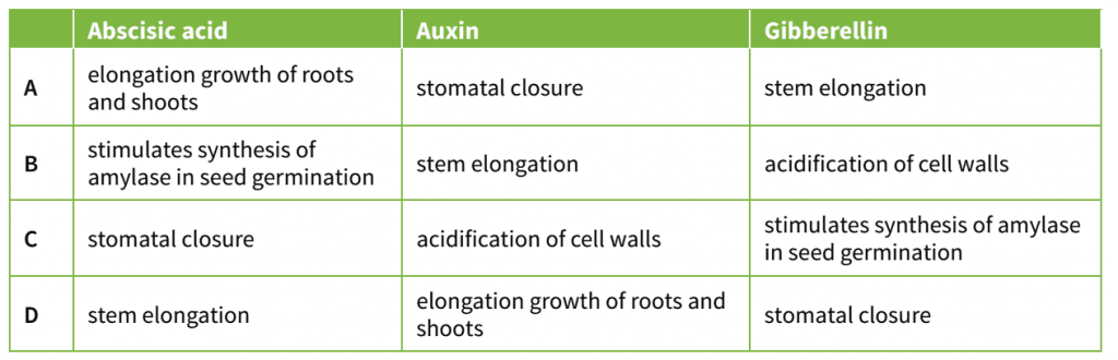
C;
5. Which statements about the concentrations of hormones in the human menstrual cycle are correct? [1]
- Shortly before ovulation, the concentration of oestrogen is high and concentration of progesterone low.
- During the last quarter of the cycle, the concentrations of oestrogen and progesterone fall.
- At the end of menstruation, the concentration of oestrogen is low but rising, and the concentration of progesterone is low.
- Just before ovulation, the concentrations of LH and FSH suddenly rise.
A 1, 2, 3 and 4
B 1, 2 and 4 only
C 2 and 3 only
D 3 and 4 only
A;
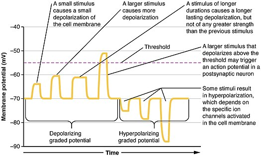
Question 6
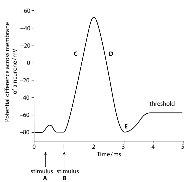
The figure shows the changes in potential difference across the membrane of a neurone over a period of time. The membrane was stimulated at time A and time B with stimuli of different intensities.
a. Stimulus B resulted in an action potential.
Describe what is occurring at C, D and E. [6]
C
sodium ions, diffuse in / enter (the neurone);
either depolarisation / described as the inside of the membrane becoming more positive or less negative;
D
potassium ions, diffuse out (from the neurone);
either repolarisation / described as the inside of the membrane becoming more negative or less positive;
E
refractory period;
hyperpolarisation occurs / inside of the membrane is more negative than resting potential;
b. Suggest why stimulus A did not result in an action potential being produced whereas stimulus B did. [2]
the generator (or receptor) potential in A was not above the threshold (potential);
in B it was above the threshold (potential);
Question 7
The electron micrograph shows parts of some myofibrils in a striated muscle that is in a relaxed state.
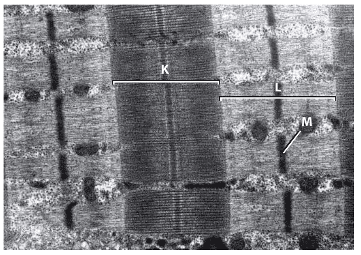
a. i Name the parts labelled K, L and M. [3]
K A band;
L I band;
M Z, line / disc;
ii. How many myofibrils are visible in the electron micrograph? Explain your answer. [2]
5;
the myofibrils are separated from each other by mitochondria;
b. i There are many glycogen granules and mitochondria visible in the electron micrograph. Explain why they are both there. [2]
glycogen granules are broken down to provide glucose for respiration;
mitochondria, carry out aerobic respiration / provide much ATP (for muscle contraction);
ii. Describe how you can tell that this electron micrograph is from relaxed muscle and not contracted muscle. [3]
there is a very wide I band;
in the I band there is no overlap between thick and thin filaments;
in contracted muscle the thin filaments would be closer together giving a thin I band;
c. The electron micrograph is magnified 20 000 times. Calculate the actual length of the sarcomere which includes the region labelled K. Give your answer in micrometres (μm). [2]

distance = 54 mm (accept 53 mm)
= 54 000 µm
so length = 54 20 000 000;
length = 2.7 µm;
Question 8
The diagrams show a sarcomere in different states of contraction.
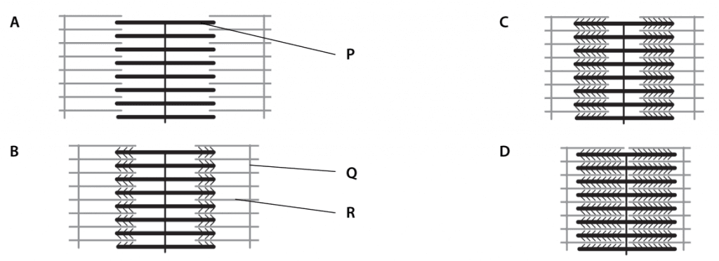
a. Name the parts labelled P, Q and R. [3]
P myosin / thick, filament;
Q actin / thin, filament;
R Z, line / disc;
b. Explain why there are no actin–myosin cross-bridges visible in diagram A. [2]
the muscle is relaxed;
the troponin and tropomyosin molecules are covering the myosin-binding sites on the, actin / thin, filaments;
myosin cannot bind to actin to form crossbridges;
c. Muscle fibres are able to contract with more force in some states of contraction than others. Suggest which of the diagrams shows the state that can develop the greatest force, and explain the reasons for your answer. [4]
diagram C;
greatest overlap between thick and thin filaments;
maximum number of cross-bridges can form between thick and thin filaments;
greatest force applied by movement of myosin heads;
in the state shown in diagram D the sarcomere cannot shorten any more;
d. Explain why the muscle shown in diagram D would not be able to contract any further. [1]
the sarcomere cannot shorten any more, without crumpling the thick and thin filaments;
the thin filaments cannot move any closer together / thick filament has reached the Z line;
e. A muscle can contract with force, but it cannot pull itself back to its original relaxed length.
i. With reference to the mechanism of muscle contraction, explain why this is so. [2]
the myosin heads can only tilt in one direction;
they are arranged so that when they do this they pull on the thin filaments in such a way that the sarcomere is shortened;
they cannot pull (or push) the thin filaments the other way;
ii Suggest how the muscle in diagram D could be returned to the state shown in diagram A. [2]
most skeletal muscles are arranged in antagonistic pairs;
contraction of an antagonist pulls on the muscle so sarcomeres lengthen;
thin filaments slide past thick filaments to give, less overlap between them / wider I band;
for example when the biceps is contracted and shortened it can be pulled back into its longer state by contraction of the
triceps muscle
Question 9
A biopsy was taken from a leg muscle of a healthy racehorse. The muscle fibres were teased apart and cross-sections were taken from one of the muscle fibres. These cross-sections were examined with a transmission electron microscope. The figure shows drawings made from three different cross-sections of a myofibril from the muscle fibre.
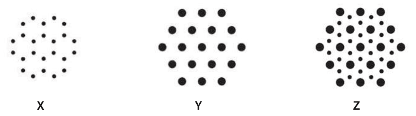
a. Explain the diff erences between the sections X, Y and Z. You may draw a labelled diagram to illustrate your answer. [4]
The diagram shows (TS) of, sarcomere / myofibril / thick and thin filaments;
X shows, actin / thin, filaments, alone; see diagram below
Y shows, myosin / thick, filaments alone; see diagram below
Z is overlap between thick and thin filaments; see diagram below
X is I band;
Z is (overlap region of) A band;
Y is H zone;
Each thick filament is surrounded by six thin filaments. The thick filaments form cross bridges with all of them.

b. The sections were taken from a relaxed muscle fibre. Suggest how the sections would appear if taken from a fibre that had contracted to its maximum extent. Explain your answer. [3]
if contracted muscle, then, no / little, I band and H zone;
so there would be no section like X and no section like Y;
thin filaments are brought closer together by action of myosin heads;
thick and thin filaments slide over each other during contraction;
c. Muscle weakness in racehorses may sometimes be related to a deficiency of calcium.
Outline the roles of calcium ions in the coordination of muscle contraction. [6]
role of calcium ions at the end of a motor neurone to max. 3:
at, neuromuscular junction / motor end plate;
voltage-gated channel proteins for calcium ions open when impulse arrives;
calcium ions enter / AW, when action potential arrives;
stimulates vesicles to, move towards / fuse with, pre-synaptic membrane;
role of calcium ions in muscle to max. 3:
impulse / action potential, in, sarcolemma / T-tubules, stimulates release of calcium ions from sarcoplasmic reticulum;
calcium ions bind to troponin;
stimulates movement of tropomyosin;
myosin binding sites on thin filaments exposed;
cross-bridges form;
when no action potential calcium ions pumped back into sarcoplasmic reticulum;
Question 10
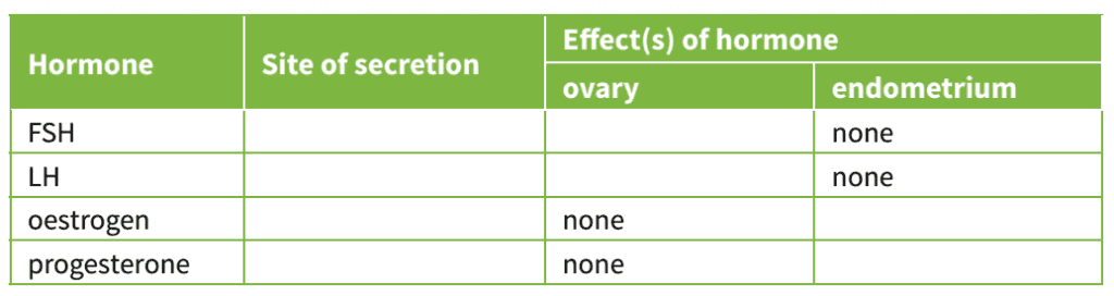
Copy and complete the table to show, for each hormone, the precise site of its secretion, and its effects on the ovary or on the endometrium of the uterus. [8]
Question 11
The Pacinian corpuscle is a type of receptor found in the dermis of the skin. Pacinian corpuscles contain an ending of a sensory neurone, surrounded by several layers of connective tissue called a capsule. The activity of a Pacinian corpuscle was investigated by inserting microelectrodes into the axon at the positions shown in the diagram below.
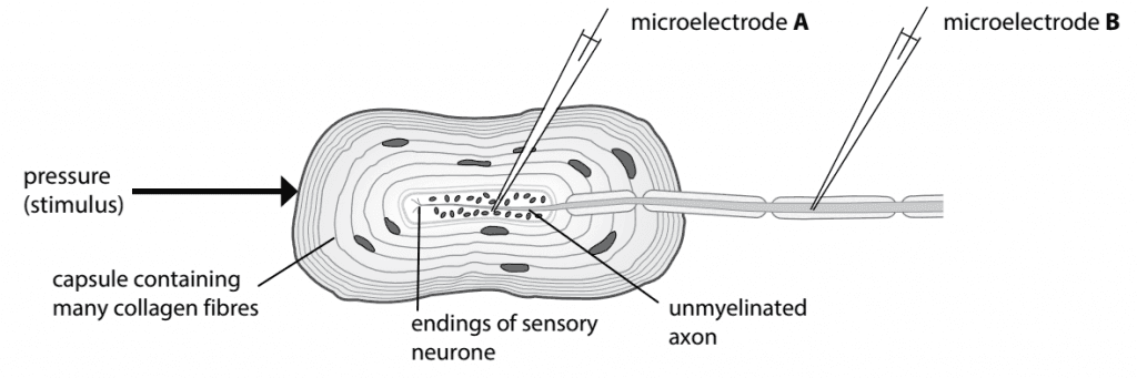
Pressure was applied to the Pacinian corpuscle and recordings made of the electrical activity in the axon at microelectrodes A and B. The results are shown in the diagram below.
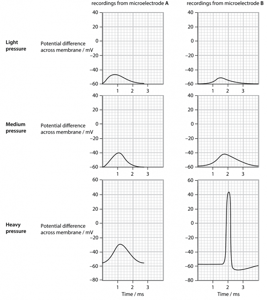
a. Suggest what happened in the unmyelinated region of the axon as pressure was applied to the Pacinian corpuscle. [4]
ref to generator / receptor potential;
pressure / stimulus, causes deformation / AW (of Pacinian corpuscle);
increased permeability to sodium ions / sodium channels open;
sodium (ions) move into, receptor / axon;
causes depolarisation / AW (e.g. potential difference changes from –ve to +ve;
b Explain the pattern of recordings from microelectrode B as the pressure applied to the corpuscle was increased. [4]
No action potential recorded with low and medium pressure / action potential recorded only with high pressure;
(no action potential at B because) depolarisation of, receptor / unmyelinated part of axon, did not reach threshold;
threshold is between medium and high pressure;
threshold is between –40 mV to –30 mV;
more depolarisation at higher pressure because more sodium ion voltage-gated channels open;
sensory neurone either conducts impulses or it does not / ref to all-or-none principle;
c Explain why sensory neurones from Pacinian corpuscles are myelinated and not unmyelinated. [3]
myelinated neurones transmit impulses much faster than unmyelinated neurones;
any suitable figures, e.g. up to 100 m s−1 for myelinated and 0.5 m s−1 for unmyelinated;
stimulation of Pacinian corpuscle might indicate damage to the skin;
may need to remove the part of the body from danger very quickly;
ref to a reflex arc in which Pacinian corpuscle provides the sensory input;
Question 12
Gibberellin is a plant growth regulator.
a. Outline the role of gibberellin in the germination of seeds such as those of wheat and barley. [5]
gibberellin secreted by embryo;
stimulates cells in the aleurone layer;
stimulates protein synthesis;
to make amylase;
to break down starch in the endosperm;
mobilises glucose;
for respiration;
to provide energy for germination;
In an investigation of the effects of gibberellin, plants of short-stemmed and long-stemmed varieties of five cultivated species were grown from seed. The young plants of each species were divided into two groups.
One group of plants was sprayed with a solution of gibberellin each day. A control group was sprayed with the same volume of water. After eight weeks, the stem length of each plant was measured and means calculated for each group of plants. A statistical test was carried out to determine whether the difference between the treatments for each species was significant.
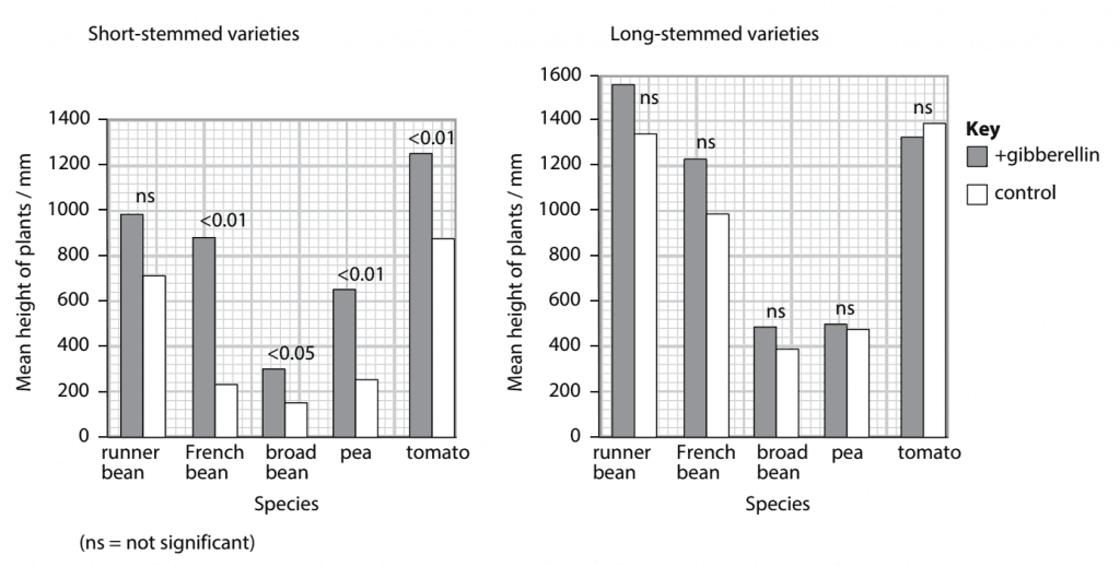
The results are shown in the figure. The p value for each species is given.
b. Using the information in the figure, describe the eff ect of adding gibberellin solution to the two varieties of the five species. [5]
gibberellin increases the mean length of the stem in the short-stemmed varieties of all species;
figures for any one species;
increase is significant in all but one species;
gibberellin increases the mean length of the stem in the long-stemmed varieties of four of the species;
not tomato;
increases are not significant;
c. Explain why the short-stemmed variety of pea showed a more significant growth in height when treated with gibberellin than the long-stemmed variety. [3]
short-stemmed plants do not make (much) gibberellin;
(because) they do not have the allele for the enzyme that makes gibberellin;
long-stemmed variety has dominant allele for gibberellin synthesis;
gibberellin supplied each day promotes growth of stems;
d. Suggest the advantages of cultivating crops of short-stemmed varieties of peas and beans rather than long-stemmed varieties. [3]
less energy used for growth of stem;
(therefore) more energy in, peas / beans / seeds;
plants do not need (as much) support;
less likely to be damaged by wind;
less plant material to harvest / less wastage at harvest;
Question 13
a. Explain how a nerve impulse is transmitted along a motor neurone. [9]
resting potential;
anything in the range –60 to –70 mV;
sodium–potassium pump uses ATP to pump
Na+ out and K+ in;
many anions inside the neurone;
resting potential is due to leakage of K+ out;
action potential is depolarisation of membrane;
up to +40 mV;
opening of voltage-gated sodium ion channels / sodium ions flow in;
closing of voltage-gated sodium ion channels;
voltage-gated potassium ion channels open;
K+ flow out;
resting potential restored;
local circuits depolarise next part of, membrane / axon;
refractory period ensures impulse does not travel backwards;
saltatory conduction in myelinated neurones;
action potential only at nodes of Ranvier;
b. Describe how an impulse crosses a synapse. [6]
action potential arrives at presynaptic membrane;
voltage-gated calcium ion channels open;
calcium ions enter to stimulate vesicles to move to membrane;
vesicles fuse with membrane / exocytosis, to release (named) neurotransmitter;
(named) neurotransmitter diffuses across (synaptic) cleft;
binds with receptor on postsynaptic membrane;
stimulates opening of sodium ion channel proteins;
sodium ions flow in through postsynaptic membrane / depolarisation of postsynaptic membrane;
Question 14
a. Describe a reflex arc and explain why such reflex arcs are important. [7]
reflex arc to max. 5:
(named) receptor cell / receptor is end of sensory neurone;
generator / receptor, potential as stimulus is above threshold;
action potential / impulses, pass along sensory neurone (in spinal nerve);
through dorsal root of spinal nerve;
into spinal cord;
synapse between sensory neurone and relay neurone;
synapse between relay neurone and motor neurone;
a synapse between sensory and motor in monosynaptic reflex such as the knee jerk reflex that has no relay neurone;
action potential / impulses, pass along motor neurone to effector;
effector / (named) muscle, contracts (to give a response);
importance to max. 2:
response to stimulus is, fast / immediate / automatic;
prevents / reduces, any damage to, (named) tissues / organs / the body;
no, decision making / thought required;
A brain not involved in coordinating the response
b. Describe the structure of a myelin sheath and explain its role in the speed of transmission of a nerve impulse. [8]
myelin sheath:
made by Schwann cells that wrap around the axon;
sheath is made of membranes that are mainly made of phospholipids;
(myelin sheath) insulates axon (membrane);
sodium and potassium ions, cannot pass through membrane that is surrounded by myelin / can only pass through membrane at nodes of Ranvier;
depolarisation (of axon membrane) only occurs at nodes of Ranvier;
local circuits / current flows, between nodes;
action potentials ‘jump’ between nodes;
this is saltatory conduction;
faster speed, of impulse transmission compared with unmyelinated neurones;
A comparison between speeds of 100 m s-1 and about 0.5 m s-1