15.04 Neuronal Impulse Transmission

Reflex Arc
- Pathway: Sensory neurone → Intermediate (relay) neurone → Motor neurone.
- Function: Provides rapid, automatic responses to stimuli (e.g., reflex actions) without involving conscious brain processing.
- Examples:
- Simple Reflexes: Withdrawal from sharp/hot objects, blinking, eye focusing, adjusting pupil size.
- Direct Reflex Arc: Certain reflexes, such as the knee-jerk, bypass the relay neurone, directly connecting sensory and motor neurone in the spinal cord for an even faster response. This motor neurone sends an impulse back to the quadriceps, causing it to contract and produce the kick. An interneuron is involved indirectly in a separate pathway that inhibits the hamstring muscles to prevent opposing movement.

Transmission of Nerve Impulses
- Nerve Impulse: Rapid electrical signal transmitted along neurones.
- Action Potential:
- Definition: Temporary reversal of membrane potential from -70 mV to +30 mV.
- Mechanism: Controlled movement of sodium (Na⁺) and potassium (K⁺) ions across the axon membrane via ion channels.
- Voltage-Gated Channels: Ion channels that open/close in response to voltage changes; critical for initiating and propagating action potentials.

Resting Potential
- Definition: Stable membrane potential when a neurone is at rest, generally between -60 mV to -70 mV.
- Maintenance Mechanisms:
- Sodium-Potassium Pump: Actively transports 3 Na⁺ out and 2 K⁺ in using ATP.
- Impermeable to Na⁺: Membrane prevents Na⁺ inflow, maintaining a negative charge inside.
- Negatively Charged Proteins: Large, negatively charged molecules inside the cell contribute to the overall negative potential.
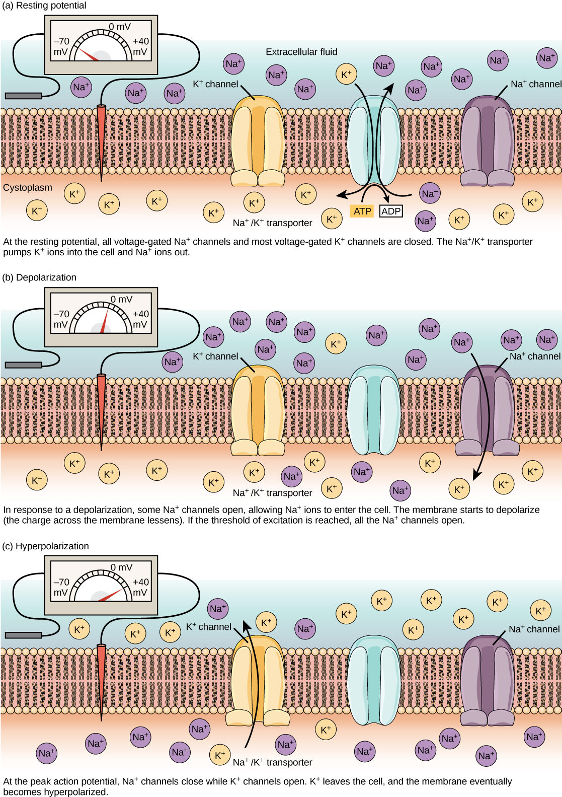
Formation of Action Potentials
- Stimulus: Opens voltage-gated Na⁺ channels, allowing Na⁺ to enter the axon, causing depolarization.
- Depolarization Threshold: When the potential reaches approximately -50 mV, a full action potential is triggered, reaching +30 mV.
- Positive Feedback: Increased Na⁺ inflow causes more Na⁺ channels to open, further depolarizing the axon.
- Repolarization:
- Na⁺ channels close; K⁺ channels open, allowing K⁺ to exit the axon, restoring a negative internal potential.
- Hyperpolarization: Brief overshoot below -70 mV before returning to the resting potential.

FIGURE: The formation of an action potential can be divided into five steps: (1) A stimulus from a sensory cell or another neuron causes the target cell to depolarize toward the threshold potential. (2) If the threshold of excitation is reached, all Na+ channels open and the membrane depolarizes. (3) At the peak action potential, K+ channels open and K+ begins to leave the cell. At the same time, Na+ channels close. (4) The membrane becomes hyperpolarized as K+ ions continue to leave the cell. The hyperpolarized membrane is in a refractory period and cannot fire. (5) The K+ channels close and the Na+/K+ transporter restores the resting potential.

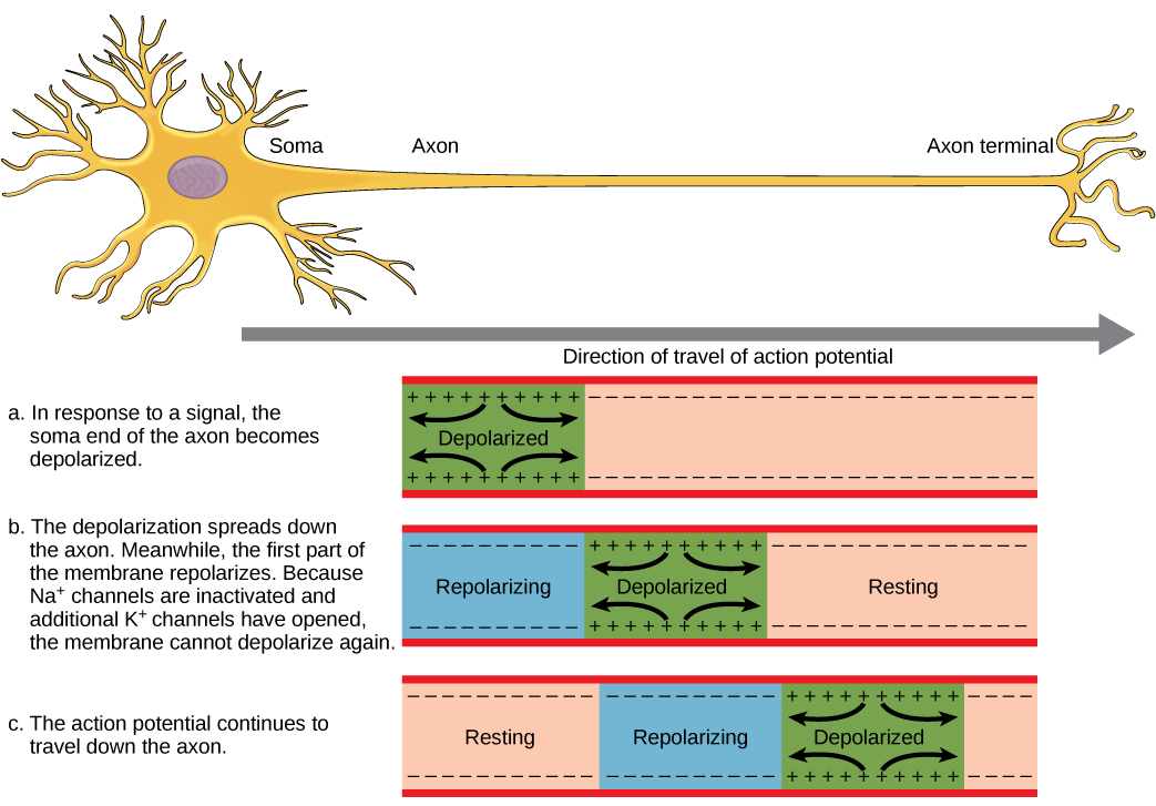
Propagation of Action Potentials
- Local Circuit: Depolarization at one point on the axon triggers depolarization in adjacent regions, creating a wave-like propagation.
- Current Flow: Moves between depolarized and resting regions, pushing the impulse forward along the axon.
- Impulse Direction: Only forward due to the refractory period (backward movement is prevented by temporarily inactive channels).
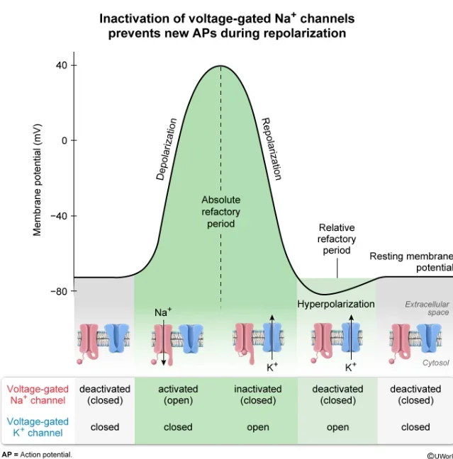
The Refractory Period
- Definition: Recovery phase following an action potential, during which the axon cannot generate a new action potential.
- Mechanism: Sodium channels behind the action potential are closed and inactive.
- Roles:
- Ensures discrete action potentials (no overlap).
- Establishes a minimum time between impulses.
- Limits impulse frequency to 200-300 per second.
- Enforces one-way direction of impulse travel along the neurone.
Information Encoding by Action Potentials
- Action Potential Consistency: Each action potential reaches a peak of +30 mV regardless of stimulus strength.
- Impulse Frequency:
- Strong Stimulus: High frequency of action potentials.
- Weak Stimulus: Lower frequency.
- Brain Interpretation: Uses frequency and the number of activated neurones to determine stimulus strength.
- Stimulus Type Identification: Based on the specific sensory neurone pathway (e.g., signals from the retina are interpreted as light).
Speed of Impulse Conduction
- Factors Affecting Speed:
- Myelin Sheath:
- Insulates axon, preventing ion exchange in myelinated regions.
- Action potentials only occur at nodes of Ranvier (gaps between myelin segments).
- Saltatory Conduction: Action potentials “jump” from node to node, increasing speed up to 100 m/s.
- Axon Diameter:
- Larger axons conduct impulses faster due to greater surface area for ion diffusion.
- In unmyelinated organisms like earthworms, large-diameter axons enhance conduction speed.

FIGURE: Nodes of Ranvier are gaps in myelin coverage along axons. Nodes contain voltage-gated K+ and Na+ channels. Action potentials travel down the axon by jumping from one node to the next.
Key Terms
- Saltatory Conduction: Rapid “jumping” of action potentials between nodes in myelinated axons, significantly speeding up impulse transmission.
- Depolarization: Phase where Na⁺ influx causes the axon’s interior to become positively charged.
- Threshold Potential: Minimum potential (~-50 mV) needed to trigger an action potential.
- Repolarization: Return to negative resting potential due to K⁺ efflux following depolarization.
- Refractory Period: Time during which a neurone is unresponsive to further stimuli, ensuring action potential discreteness and unidirectional flow.
Practise Questions
Test 1
1. What is the correct pathway of a reflex arc?
2. What is the primary function of a reflex arc?
3. Which component is bypassed in a direct reflex arc, allowing for a faster response?
4. What triggers the formation of an action potential in a neurone?
5. During repolarization, which ions exit the neurone to restore the resting potential?
6. What ensures that action potentials travel only in the forward direction along the neurone?
7. What is the role of the sodium-potassium pump in maintaining the resting potential?
8. What is saltatory conduction?
9. Why do larger-diameter axons conduct impulses faster?
10. What happens during the refractory period of a neurone?
Correct Answers: 0%
Test 2
1. What is the correct pathway of a reflex arc?
2. What is the primary function of a reflex arc?
3. Which component is bypassed in a direct reflex arc, allowing for a faster response?
4. What triggers the formation of an action potential in a neurone?
5. During repolarization, which ions exit the neurone to restore the resting potential?
6. What ensures that action potentials travel only in the forward direction along the neurone?
7. What is the role of the sodium-potassium pump in maintaining the resting potential?
8. What is saltatory conduction?
9. Why do larger-diameter axons conduct impulses faster?
10. What happens during the refractory period of a neurone?
Correct Answers: 0%
Test 3
1. What occurs during the refractory period of a neurone?
2. What is the primary role of the nodes of Ranvier in myelinated axons?
3. What defines the depolarization threshold in a neurone?
4. How does myelination affect the speed of nerve impulse conduction?
5. What initiates repolarization during an action potential?
6. What is the main purpose of the sodium-potassium pump in neurones?
7. What characterizes saltatory conduction in myelinated axons?
8. How does axon diameter influence the speed of impulse conduction?
9. What ensures the unidirectional flow of nerve impulses along an axon?
10. How is stimulus strength encoded by neurones?
Correct Answers: 0%
Test 4
1. In a polysynaptic reflex arc, which neuron type is introduced to allow for integration and modulation of the reflex?
2. What neurotransmitter is primarily responsible for excitatory synaptic transmission at the neuromuscular junction?
3. How does the all-or-none principle apply to action potentials in neurones?
4. Which of the following best describes the role of interneurones in the central nervous system?
5. During an action potential, what is the primary function of voltage-gated potassium (K⁺) channels?
6. Which phase of the action potential is characterized by the temporary overshoot below the resting membrane potential?
7. What mechanism ensures that an action potential propagates only in the forward direction along an axon?
8. In the context of reflex arcs, what is the primary difference between monosynaptic and polysynaptic reflexes?
9. How does the frequency of action potentials relate to stimulus intensity in neuronal signaling?
10. What role do neurotransmitters play in the transmission of nerve impulses at chemical synapses?
Correct Answers: 0%