7.04 Biological Drawings
Microscopy Drawing Guide
1. Introduction
Microscopy drawings are essential for accurately representing biological specimens observed under a microscope. They are divided into two main types: Low Power Drawings and High Power Drawings. Understanding the distinct purposes and guidelines for each type ensures clear and effective scientific illustrations.
2. Types of Microscopy Drawings
A. Low Power Drawings
Guidelines:
- Identify the Tissue:
- For low power drawings, you just want to have an overall outline of the different tissues.
- If it helps, use high power magnification to identify different tissues.
- Completely enclose each tissue with clear boundary lines.
- Simplify Representation:
- Do Not draw individual cells; focus on tissue distribution.
- Avoid filling spaces with cells to prevent gaps between tissues.
- Accuracy:
- Represent tissue distribution based on observation, not textbook images or what you think it should look like.
- Use Representative Portions:
- For symmetrical structures, draw a representative section (e.g., half of a transverse section of root or stem).
- For leaves, include half a midrib and a small portion of the adjacent lamina.
- Scale Appropriately:
- Ensure the drawing occupies at least half of the designated page space.
- Enlarge all parts of drawing equally (keep the same uniform ratio and shape of the cells/tissue, just drawn larger).
- Drawing Area:
- Utilize the allocated space efficiently, covering around half to two-thirds of the provided area (rather draw large than too small).
- Avoid drawing over text or page margins.
B. High Power Drawings
Guidelines:
- Select Representative Cells:
- Draw only a few adjacent cells (typically 2-3) to demonstrate cell structure and arrangement.
- If cells are similar, three cells are often sufficient.
- If they ask you to draw cells that are touching each other, make sure all 3 cells touch each of the other cells.
- Simplify Cellular Components:
- Do Not shade nuclei or nucleoli; draw only their outlines.
- Drawing Size:
- Ensure the drawing covers around two-thirds of the allocated space for clarity.
- Avoid:
- Inaccurate representation of cells.
3. Essential Tools
- Sharp HB or H Pencil: For clear, precise pencil lines.
- Eraser: To correct mistakes without damaging the paper.
- Ruler: For drawing straight lines, measuring scales, and underlining headings.
- Plain Paper: Use high-quality, unlined paper for clarity.
- Pen: For labelling and headings only (do not use for the main drawing).
4. General Drawing Quality Standards
- Line Usage:
- Use clear, continuous pencil lines for the main drawing.
- Do Not use a pen for the main drawing.
- No Shading:
- Avoid shading; only draw the outlines of objects, regardless of their actual darkness.
- Proportions:
- Ensure accurate proportions based on direct observation, not textbook images.
- Scale Appropriately:
- Draw the specimen to occupy at least half of the drawing page.
- Drawing Area:
- Make the drawing large enough to cover approximately two-thirds of the designated space.
- Draw within the provided space, avoiding overlapping text or page margins.
- Accuracy and Clarity:
- Draw what you see under the microscope, not what you expect based on prior knowledge.
- Maintain precise and neat lines using a sharp pencil and ruler.
5. Labelling Standards
- Writing Tools:
- Use pen for all labels and headings.
- Use pencil for label lines to allow corrections and maintain clarity.
- Headings:
- Each drawing should have a heading written in pen.
- Underline the heading using a ruler and pen for neatness.
- Label Placement:
- Labels should be horizontal and aligned neatly, typically on the right side of the drawing.
- Arrange labels below each other to avoid clutter.
- Label Lines:
- Use straight lines drawn with a ruler.
- Do Not use arrowheads; label lines should be straight without any arrowheads.
- Annotations:
- Add explanatory notes if necessary to clarify specific features.
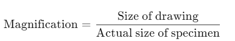
6. Magnification and Scale
A. Calculating Magnification
- Formula:

- Example Calculation:
- If magnification is 400× and a scale line in the drawing is 40 mm: 40 mm (drawing) = 400 μm (specimen)
- Steps:
- Write the formula for magnification.
- Substitute the given values with appropriate units.
B. Scale Line
- Purpose: Represents the real size of structures within the specimen.
- Placement: Typically placed at the bottom of the drawing.
- Example:
- At 400× magnification, a 40 mm scale line in the drawing corresponds to 400 μm in the specimen.
7. Common Errors in Drawings
- Arrowheads on Label Lines: Label lines should be straight without any arrowheads.
- Use of Shading: Shading is not allowed in scientific drawings; only outlines should be drawn.
- Overlapping or Disconnected Lines: Ensure lines are clear and connected to avoid confusion.
- Incorrect Proportions: Maintain accurate proportions based on observation to reflect true tissue distribution and cell structures.
- Uneven or Crossed Label Lines: Labels should be neatly aligned without overlapping lines.
- Drawing with a Pen: All main drawings should be done in pencil; pens are only for labels and headings.
- Labelling Mistakes: Avoid using pens for label lines or writing labels in pencil.
8. Examples for Reference: High Power Drawings
A. Mouse Pancreas Section
- Figures A:
- Display photomicrographs alongside low and high power drawings.

- Examples (of drawings submitted by students):

Critique:
Good:
- Both drawings have a heading.
- Both indicate the magnification.
Bad:
- The overall shape is different between the two examples. The drawing on the left is the more accurate shape.
- The label lines are not horizontal.
- The labels on the right specifically do not line up below each other.
- The drawing on the right has some thicker and thinner lines (aka, shading).
- In both drawings, it seems that the drawing and the writing is in pen or pencil.
B. Beech Leaf Sections (Fagus)
- Figures B:
- Show photomicrographs and corresponding low and high power drawings.
- Compare leaves from sunny and shaded conditions.

Examples (of drawings submitted by students):

Critique:
Good:
- Both drawings have a heading.
- Both indicate the magnification.
Bad:
- In both drawings, it seems that the drawing and the writing is in pen or pencil.
- The drawing on the left is definitely too small.
- Both drawings seemed to have used shading and made the nuclei of the cells darker (all lines and structures should have the same thickness of pencil lines).
- The spacing between the cells on the drawing on the left seems to be off. The drawing on the right seems to have the right amount/proportion of spacing.
C. Islet of Langerhans from the pancreas


Examples (of drawings submitted by students):


Critique:
Good:
- The general proportion of the cells seems to be ok.
Bad:
- Read the comments on the side of the drawing for more information.
9. Examples for Reference: Low Power Drawings
D Transverse section of the lamina of a shade leaf of beech (Fagus)

Example (of drawings submitted by students):

E Transverse section of the lamina of a sun leaf of beech


F Transverse section of a dicot stem
Example (of drawings submitted by students):


The drawing made by the student on the right is far more accurate than the one on the left.
Other Examples:


11. Summary
- Practice and Accuracy: Regular practice and a focus on accurate observation enhance drawing skills and the ability to effectively represent microscopic structures.
- Low Power Drawings: Focus on accurately mapping the distribution and boundaries of tissues within an organ without depicting individual cells. Serve as a structural overview.
- High Power Drawings: Highlight detailed cell structures and their arrangements, complementing low power drawings for a comprehensive understanding.
- Essential Tools and Quality Standards: Utilize the correct tools and adhere to quality guidelines to ensure clear, accurate, and professional scientific drawings.
- Labelling and Scale: Maintain precise and neat labeling standards, and accurately represent magnification and scale to reflect true specimen dimensions.
- Avoid Common Errors: Be mindful of common pitfalls such as shading, incorrect proportions, and improper labeling to maintain the integrity of scientific drawings.
Practice Questions
Question 1
Define low power and high power microscopy drawings and explain their distinct purposes. (5 marks)
Mark Scheme:
- Definition of Low Power Drawings:
- Provide an overall outline of different tissues without depicting individual cells. (1 mark)
- Definition of High Power Drawings:
- Focus on detailed cell structures and their arrangements, typically showing 2-3 adjacent cells. (1 mark)
- Purpose of Low Power Drawings:
- Serve as a structural overview, showing the organization of tissues within an organ. (1 mark)
- Purpose of High Power Drawings:
- Highlight cellular morphology and arrangement, complementing low power drawings for comprehensive understanding. (1 mark)
- Distinct Features:
- Low power emphasizes tissue distribution; high power emphasizes cell details without shading. (1 mark)
Question 2
List and describe three guidelines for creating accurate low power microscopy drawings. (6 marks)
Mark Scheme:
- Identify the Tissue:
- Outline different tissues with clear boundary lines based on observations. (1 mark)
- Simplify Representation:
- Do not draw individual cells; focus on tissue distribution without filling spaces with cells. (1 mark)
- Use Representative Portions:
- For symmetrical structures, draw a representative section (e.g., half of a transverse section). (1 mark)
- Scale Appropriately:
- Ensure the drawing occupies at least half of the designated page space, enlarging all parts uniformly. (1 mark)
- Maintain Drawing Area:
- Utilize the allocated space efficiently, covering around half to two-thirds of the provided area without overlapping text or margins. (1 mark)
- Accuracy:
- Represent tissue distribution based on direct observation, not textbook images or assumptions. (1 mark)
Question 3
Explain the importance of using a sharp HB or H pencil in microscopy drawings. (5 marks)
Mark Scheme:
- Clarity and Precision:
- Sharp pencils produce clear, precise lines essential for accurate representation. (1 mark)
- Detail Representation:
- Allows for the depiction of fine details in cell structures and tissues without smudging. (1 mark)
- Ease of Correction:
- Pencil lines can be easily erased and corrected without damaging the paper. (1 mark)
- Consistency:
- Provides consistent line quality, maintaining uniformity throughout the drawing. (1 mark)
- Professional Appearance:
- Enhances the neatness and readability of scientific illustrations, adhering to quality standards. (1 mark)
Question 4
Describe the role of stomata in leaves and how they should be represented in high power microscopy drawings. (5 marks)
Mark Scheme:
- Function of Stomata:
- Facilitate gas exchange, allowing the intake of CO₂ and release of O₂ during photosynthesis. (1 mark)
- Structural Representation:
- Draw pores (openings) on the leaf surface where stomata are located. (1 mark)
- Arrangement:
- Represent stomata as small, evenly spaced openings without shading nuclei or nucleoli. (1 mark)
- Labeling:
- Clearly label stomata using horizontal label lines without arrowheads. (1 mark)
- Accuracy:
- Ensure the size and placement of stomata reflect their actual distribution as observed under high power magnification. (1 mark)
Question 5
List the essential tools required for creating microscopy drawings and briefly state their purposes. (6 marks)
Mark Scheme:
- Sharp HB or H Pencil:
- For drawing clear, precise lines in the main illustration. (1 mark)
- Eraser:
- To correct mistakes without damaging the paper. (1 mark)
- Ruler:
- For drawing straight lines, measuring scales, and underlining headings. (1 mark)
- Plain Paper:
- Use high-quality, unlined paper to ensure clarity and prevent distortion of lines. (1 mark)
- Pen:
- For labeling and headings only, ensuring they are neat and consistent. (1 mark)
- Additional Tools (optional):
- Compass for drawing circles, if necessary. (1 mark)
Question 6
Explain how to correctly label a microscopy drawing according to the guidelines. (5 marks)
Mark Scheme:
- Writing Tools:
- Use a pen for all labels and headings, and a pencil for label lines. (1 mark)
- Headings:
- Each drawing should have a heading written in pen and underlined using a ruler and pen for neatness. (1 mark)
- Label Placement:
- Place labels horizontally and aligned neatly on the right side of the drawing. Arrange labels below each other to avoid clutter. (1 mark)
- Label Lines:
- Use straight lines drawn with a ruler to connect labels to structures without using arrowheads. (1 mark)
- Annotations:
- Add explanatory notes if necessary to clarify specific features, ensuring they do not clutter the drawing. (1 mark)
Question 7
Describe the process of adding a scale line to a microscopy drawing and its significance. (5 marks)
Mark Scheme:
- Placement of Scale Line:
- Typically placed at the bottom of the drawing for easy reference. (1 mark)
- Representation of Real Size:
- The scale line represents the actual size of structures based on the magnification used. (1 mark)
- Calculation Formula:
- Real Size = Drawing Size / Magnification. (1 mark)
- Example Application:
- At 400× magnification, a 40 mm scale line in the drawing corresponds to 0.1 mm or 100 μm in the specimen. (1 mark)
- Purpose of Scale Line:
- Ensures the drawing accurately reflects the true dimensions of the specimen, aiding in measurement and analysis. (1 mark)
Question 8
Identify three common errors in microscopy drawings and provide solutions to avoid them. (6 marks)
Mark Scheme:
- Shading:
- Error: Shading cell structures to indicate depth or density.
- Solution: Only draw outlines of cells and tissues without shading. (1 mark)
- Incorrect Proportions:
- Error: Distorting the size and shape of tissues or cells based on expectations rather than observation.
- Solution: Ensure accurate proportions by closely following the microscope image and measuring if necessary. (1 mark)
- Use of Pen for Main Drawing:
- Error: Using a pen for the main drawing instead of a pencil, making corrections difficult.
- Solution: Use a sharp pencil for the main drawing and reserve pens for labels and headings only. (1 mark)
- Imbalanced Label Lines:
- Error: Drawing uneven or crossed label lines that clutter the drawing.
- Solution: Use straight, horizontal lines for labels and arrange them neatly below each other to maintain clarity. (1 mark)
- Overcrowding the Drawing Area:
- Error: Drawing too many details or covering text and margins with the specimen.
- Solution: Simplify the drawing by focusing on key structures and utilize the allocated space efficiently without overlapping text or margins. (1 mark)
- Inconsistent Line Thickness:
- Error: Varying line thickness, which can misrepresent the actual structure.
- Solution: Maintain uniform line thickness throughout the drawing to ensure consistency and accuracy. (1 mark)
Question 9
Explain the steps to prepare samples for microscopy drawing and the importance of balancing centrifuge tubes. (6 marks)
Mark Scheme:
- Prepare Samples:
- Place the sample in centrifuge tubes and ensure all tubes have the same volume and are balanced in weight. (1 mark)
- Load the Centrifuge:
- Arrange the tubes opposite each other in the rotor to maintain balance. (1 mark)
- Set Parameters:
- Select the appropriate speed (RPM) and time based on the protocol or experiment requirements. (1 mark)
- Start the Centrifuge:
- Close the lid securely and start the machine, allowing it to spin for the set duration. (1 mark)
- Wait for Completion:
- Allow the centrifuge to complete the spin and stop completely before opening the lid to ensure safety. (1 mark)
- Retrieve Samples Carefully:
- Carefully remove the tubes, avoiding mixing the separated layers of pellet and supernatant. (1 mark)
Question 10
Discuss the role of the endodermis in dicotyledonous roots and how it regulates water and mineral movement. (5 marks)
Mark Scheme:
- Definition and Location:
- The endodermis is a single layer of cells located just inside the epidermis and surrounding the vascular tissue in dicot roots. (1 mark)
- Casparian Strip:
- The endodermal cells possess a Casparian strip, a band of suberin and lignin that blocks the passive flow of water and solutes through the cell walls. (1 mark)
- Selective Absorption:
- Forces water and dissolved minerals to pass through the plasma membranes of endodermal cells, allowing the plant to regulate and selectively absorb nutrients. (1 mark)
- Barrier Function:
- Acts as a barrier to prevent the backflow of substances, ensuring that minerals and water are directed into the xylem for transport. (1 mark)
- Regulation of Ion Uptake:
- Helps in maintaining ion balance and prevents harmful substances from entering the vascular system. (1 mark)