5.04 Mitosis
Mitosis (Nuclear Division): Introduction
- Definition: Mitosis is a type of cell division in which a single parent cell divides to produce two genetically identical daughter cells, each containing the same number of chromosomes as the parent cell.
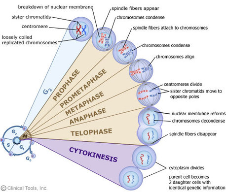
- Mitosis starts after passing the G2 checkpoint.
- Mitosis divides the nucleus into two genetically identical nuclei through four key stages: Prophase, Metaphase, Anaphase, Telophase.
- After DNA replication in the S phase, each chromosome consists of two chromatids (in other words, 46 chromatin were “copied” to form 92 chromatids held together by a centromere. From this point we refer to each of these daughter chromatin strands as chromatids. These chromatids will condense during prophase to form the X like structures that we call chromosomes. .
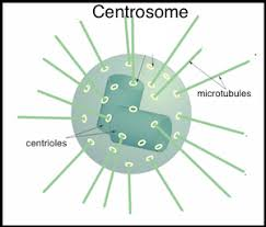
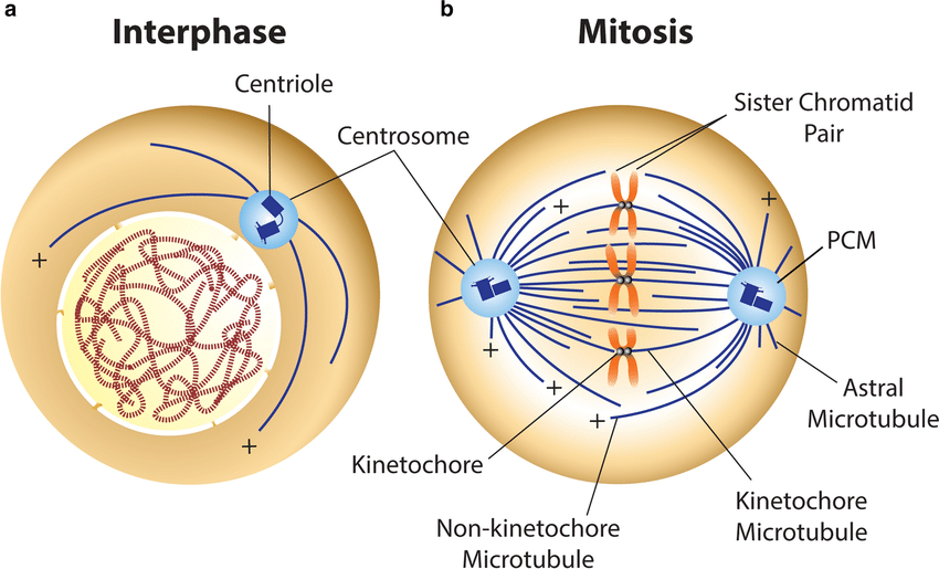
Centrioles
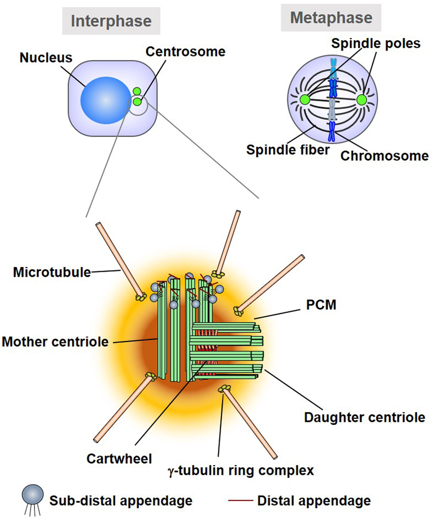
Definition of Centrioles:
- Centrioles are cylindrical structures made of microtubules, found in the centrosome (the microtubule-organizing center).
Role in Mitosis:
- Formation of Spindle Fibers: Centrioles help organize the mitotic spindle, a structure that separates chromosomes during mitosis.
- Aster Formation: They produce asters (star-like microtubule arrays) that anchor the spindle apparatus to the cell membrane.
- Chromosome Alignment: The spindle fibers, emanating from centrioles, attach to kinetochores on chromosomes, ensuring they align at the metaphase plate.
- Chromosome Separation: During anaphase, spindle fibers pull sister chromatids apart toward opposite poles of the cell.
- Ensuring Even Distribution: By organizing the spindle, centrioles ensure equal distribution of genetic material to the daughter cells.
Movement:
- Centrioles replicate during the S phase and migrate to opposite poles of the cell during prophase, positioning the spindle apparatus.
Note:
- Centrioles are essential in animal cells but are absent in most plant cells, which still form a spindle using other microtubule-organizing centers.
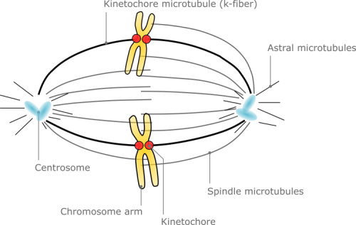
Spindle:
What is the Spindle?
- Structures made of microtubules.
- Forms during mitosis to separate chromosomes into daughter cells.
Functions of the Spindle:
- Chromosome Alignment: Ensures chromosomes align at the metaphase plate.
- Chromosome Separation: Pulls sister chromatids apart during anaphase.
- Equal Division: Guarantees accurate genetic material distribution.
Formation of the Spindle:
- Arises from the centrosomes, which include centrioles in animal cells.
- Microtubules extend outward from the centrosomes, forming the spindle apparatus.
Relation to Centrioles:
- Centrioles in the centrosomes organize and anchor the spindle fibers.
- They migrate to opposite poles during prophase, ensuring proper spindle orientation.
- Spindle fibers attach to chromosomes at the kinetochores, facilitating their movement.
Key Phases in Mitosis Involving the Spindle:
- Prophase: Spindle formation begins.
- Metaphase: Spindle fibers align chromosomes at the center.
- Anaphase: Spindle pulls chromatids apart to opposite poles.
- Telophase: Spindle disassembles as the cell prepares for division.
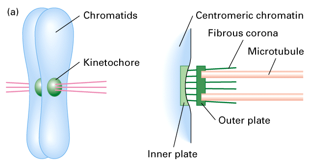
Kinetochores:
- Kinetochores are protein structures at the centromere allow spindle fibers to attach and pull chromatids toward poles.
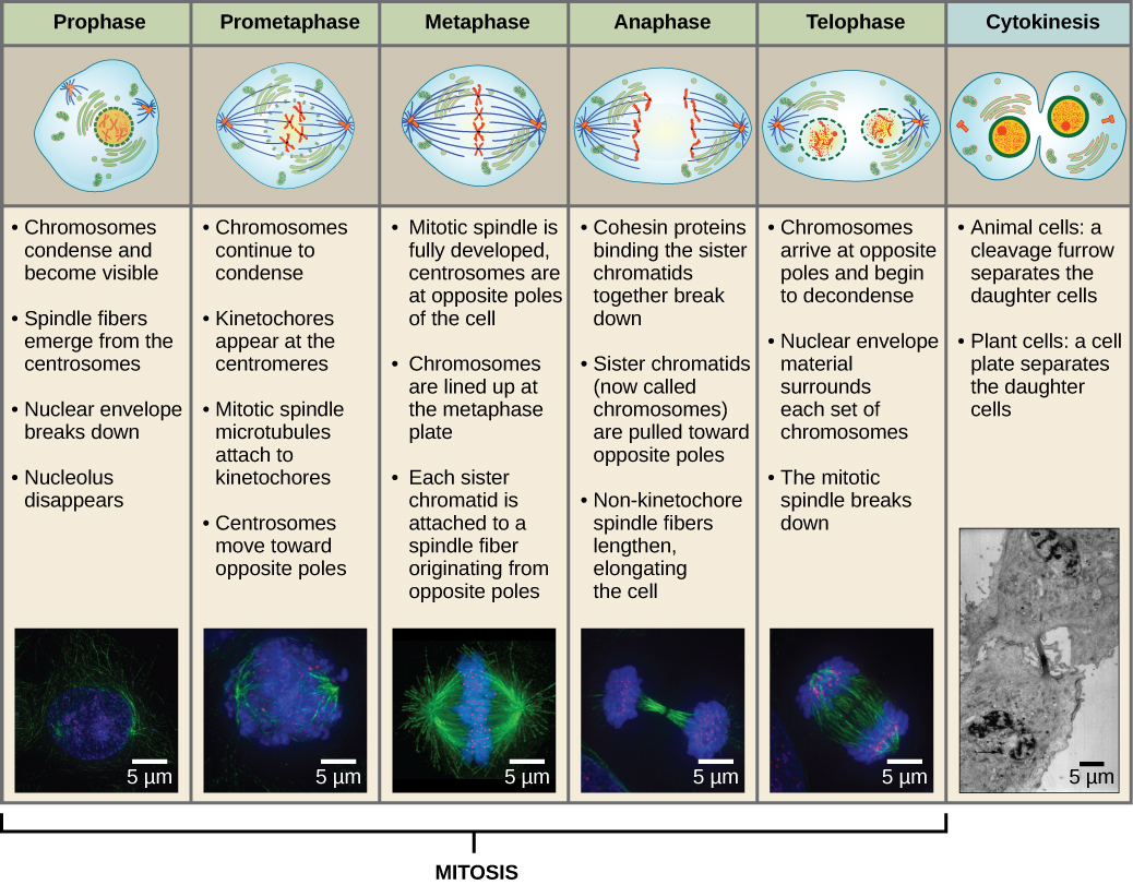
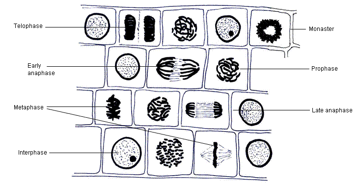
Mitosis (Nuclear Division): Phases

1. Prophase
1.1 Early Prophase
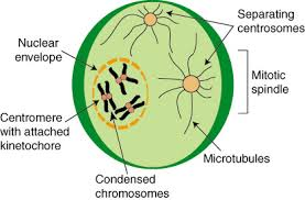
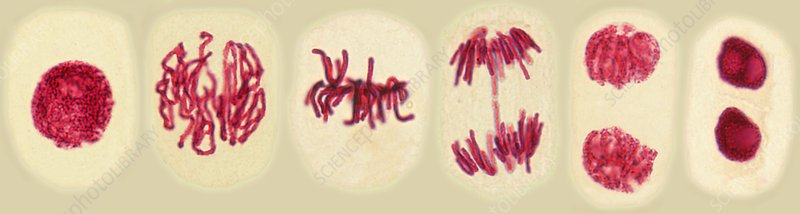
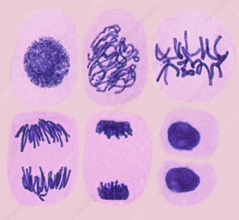
1.1.1 Chromatin Condensation:
- Chromatin fibers condense into visible chromosomes.
- Each chromosome consists of two sister chromatids joined at the centromere.
1.1.2 Nucleolus Disassembly:
- The nucleolus becomes less distinct and eventually disappears.
1.1.3 Centrosome Migration:
- Centrosomes (containing centrioles in animal cells) begin migrating to opposite poles of the cell.
1.1.4 Spindle Formation:
- Microtubules from centrosomes start forming the spindle apparatus.
1.1.5 Aster Formation:
- Star-like arrays of microtubules (asters) radiate outward from each centrosome to stabilize their position.
1.2 Late Prophase (Prometaphase)
1.2.1 Nuclear Envelope Breakdown:
- The nuclear envelope fully disintegrates, creating access between the cytoplasm and the chromosomes.
1.2.2 Chromosome Visibility:
- Chromosomes become fully visible as condensed, discrete structures.
1.2.3 Kinetochore Formation:
- Protein complexes called kinetochores form at the centromeres of each chromosome.
1.2.4 Spindle Fiber Interaction:
- Spindle fibers attach to the kinetochores, linking chromosomes to the spindle apparatus.
- Non-kinetochore spindle fibers extend to overlap at the cell’s equator, aiding cell structure.
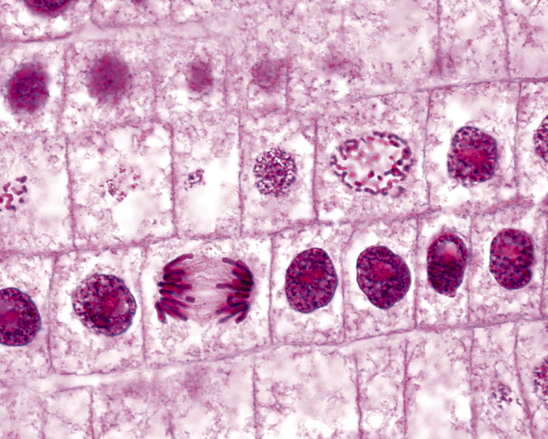
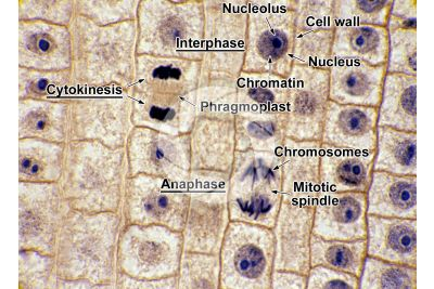
- How it looks like under a microscope:
- Chromosomes:
- Appear as dark, thread-like structures inside the nucleus, distinct from the chromatin of interphase.
- Nucleus: Nuclear envelope starts to disappear, making the boundary less distinct.
- Cytoplasm: Centrosomes may become visible as small dots near opposite ends of the cell (in animal cells).
- Chromosomes:
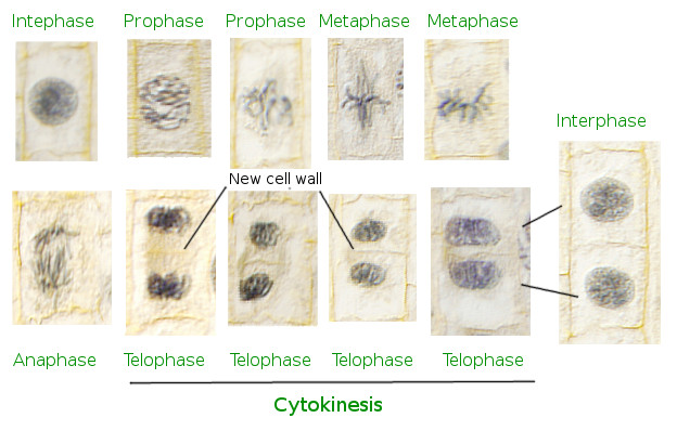
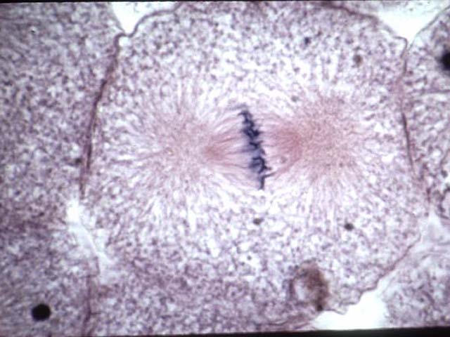
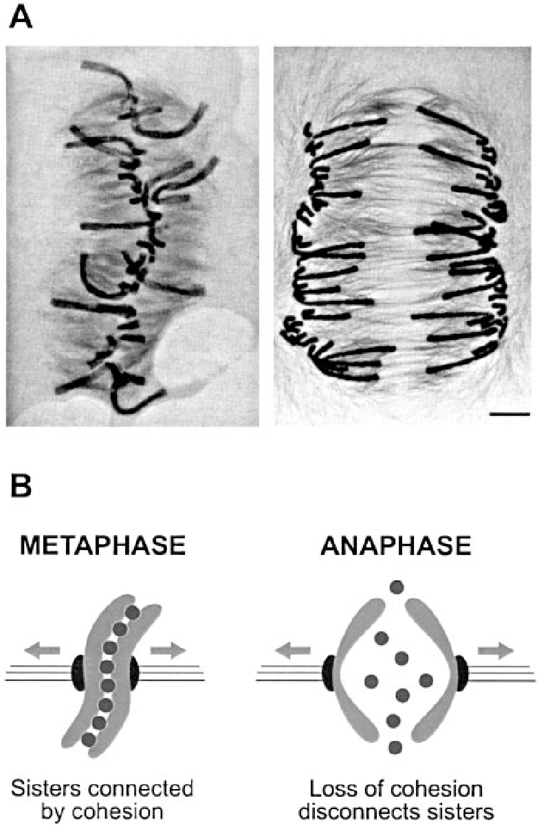
2. Metaphase
- The nuclear envelope disappears.
- The centriole pairs are at the poles.
- The spindle is completely formed.
- The chromosomes continue to condense.
- The spindle fibres attach to the
centromeres of the chromosomes. - The spindle fibres pull on the centromeres,
arranging them on the equator.
- How it looks like under a microscope:
- Chromosomes:
- Clearly visible and aligned in a single line at the center of the cell.
- Nucleus: No nuclear envelope; chromosomes are free in the cytoplasm.
- Spindle Apparatus: May appear faintly as lines extending from poles to chromosomes.
- Chromosomes:
3. Anaphase
- The links between sister chromatids break.
- The centromeres of sister chromatids
move apart, pulled by the spindle fibres.
- How it looks like under a microscope:
- Chromosomes:
- Appear as V-shaped structures moving toward opposite poles.
- Clear Division: Two groups of chromatids are visibly separating, with a clear gap between them.
- Spindle Fibers: Sometimes faintly visible, connecting chromatids to poles.
- Chromosomes:
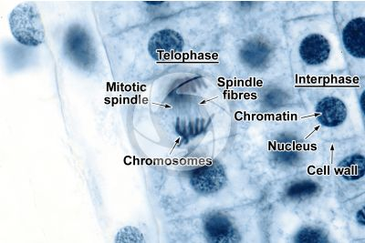
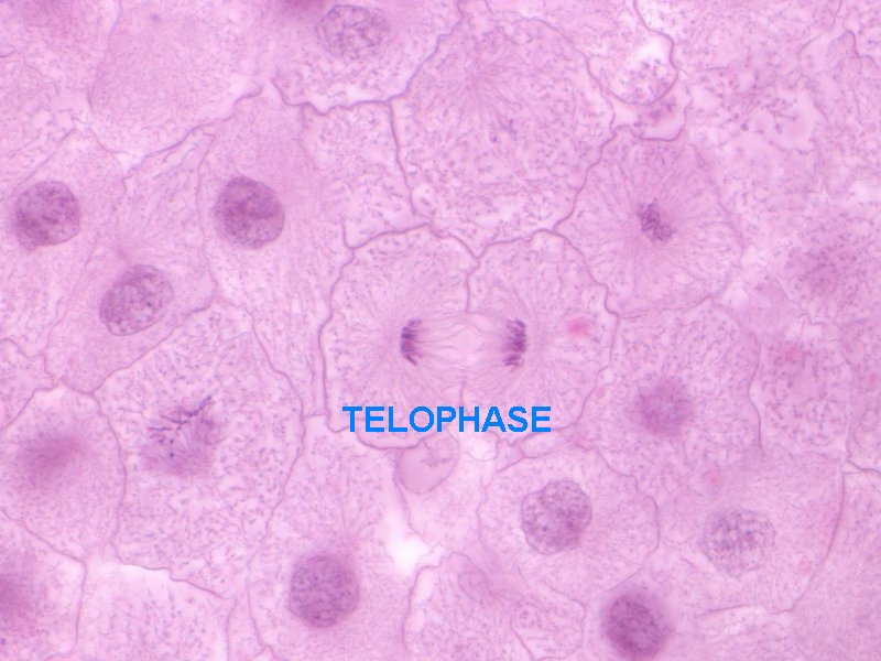
4. Telophase
- Sister chromatids (now effectively separate
chromosomes) reach opposite poles. - The chromosomes decondense.
- Nuclear envelopes begin to form around
the chromosomes at each pole. - The spindle disappears.
- How it looks like under a microscope:
- Chromosomes:
- Begin to decondense, becoming less visible.
- Nuclei:
- Two newly formed nuclei are visible, each surrounded by a distinct nuclear envelope.
- Cytoplasm:
- Early signs of cytoplasmic division (cleavage furrow or cell plate) may be visible.
- Chromosomes:
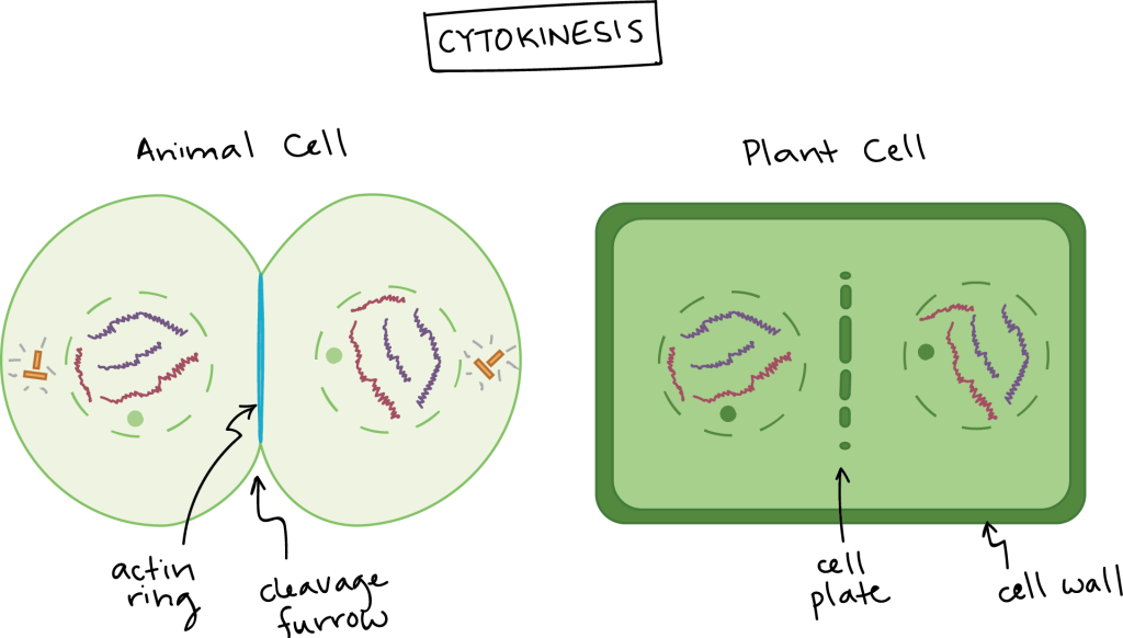
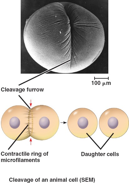
3. Cytokinesis (Cytoplasmic Division)
- The cell divides into two daughter cells, either by:
- infolding of the cell surface membrane in
animal cells, - or by the formation of a new
cell wall and cell surface membrane in
plants
- infolding of the cell surface membrane in
- How it looks like under a microscope:
- Animal Cells:
- Look for a cleavage furrow: An indentation in the cell membrane at the center.
- The cell appears to be “pinching apart” into two.
- Plant Cells:
- Look for a cell plate in the center of the cell, which will develop into the cell wall.
- The cell plate appears as a straight, dark line dividing the cytoplasm.
- Animal Cells:

MITOSIS SUMMARY
Key Differences in Microscope Slide Views Across Stages
| Stage | Nuclear Features | Chromosome Appearance | Cytoplasmic Features | Chromatid/Chromosome Count |
|---|
| Interphase | Intact nuclear membrane; diffuse chromatin | Not visible | Normal cytoplasm | Chromosomes: 46 individual chromosomes initially, replicating to form 46 duplicated chromosomes (92 chromatids). |
| Prophase | Fragmenting nuclear envelope; condensing chromosomes | Condensed, thread-like structures | Centrosomes moving to opposite poles | Chromosomes: 46 duplicated chromosomes (92 chromatids). |
| Metaphase | No nuclear envelope; spindle fibers present | Aligned at the cell’s equator | Spindle fibers attach to centromeres | Chromosomes: 46 duplicated chromosomes (92 chromatids). |
| Anaphase | No nuclear envelope | Chromatids separating (V-shaped) | Spindle fibers pulling chromatids apart | Chromosomes: 92 individual chromosomes (as chromatids separate into single chromosomes). |
| Telophase | Reforming nuclear envelope; nuclei forming | Decondensing into chromatin | Early cleavage furrow or cell plate | Chromosomes: 46 individual chromosomes in each newly forming nucleus. |
| Cytokinesis | Two separate nuclei | Not visible | Cleavage furrow or cell plate visible | Chromosomes: 46 individual chromosomes per daughter cell. |
Observing the Cell Cycle Under a Microscope
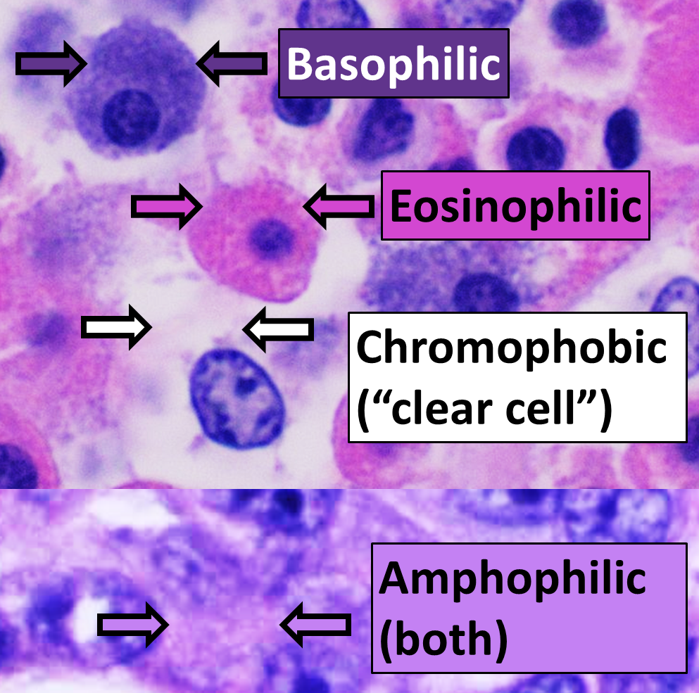
- Staining:
- Use dyes like hematoxylin and eosin (H&E), Giemsa, or DAPI to improve visibility of DNA and chromosomes.
- Tissue Type:
- Look at tissues with rapid cell division, such as the root tips of plants or intestinal lining in animals.
- Magnification:
- Higher magnification (400x or more) is typically needed to distinguish individual chromosomes.
Mitotic Checkpoint in Mitosis
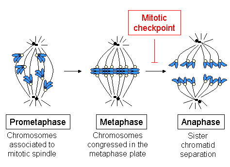
What is the Mitotic Checkpoint?
- A control mechanism in mitosis that ensures proper chromosome separation.
Purpose of the Checkpoint:
- Prevents errors in chromosome segregation.
- Ensures all chromosomes are correctly attached to spindle fibers at their kinetochores before anaphase begins.
Key Phase for the Mitotic Checkpoint:
- Functions during metaphase, just before anaphase.
How the Checkpoint Works:
- Monitors tension and attachment of spindle fibers to kinetochores.
- If errors are detected (e.g., unattached or misaligned chromosomes), the checkpoint delays progression to anaphase.
Clinical Relevance:
- Checkpoint failure can lead to genetic instability, associated with conditions like cancer.
Importance of Mitosis
- Growth: Allows multicellular organisms to grow from a single zygote, ensuring each cell has identical genetic information.
- Cell Replacement & Tissue Repair: Replaces dead or damaged cells, especially rapid in tissues like skin and the gut lining.
- Asexual Reproduction: Produces genetically identical offspring in organisms such as Amoeba and through vegetative propagation in plants.
- Immune Response: Cloning of B- and T-lymphocytes during immune responses is dependent on mitosis.
Practice Questions 1
1.Define mitosis and the cell cycle.
- Mitosis: Division of a nucleus into two identical daughter nuclei.
- Cell cycle: Sequence of events from one cell division to the next, including interphase, mitosis, and cytokinesis.
2.Human cells contain 46 chromosomes. In the mitotic cell cycle of a human cell:
- a. Number of chromatids as the cell enters mitosis?
- Answer: 92 chromatids (each chromosome has replicated to form two sister chromatids).
- b. Number of DNA molecules?
- Answer: 92 DNA molecules (one per chromatid).
- c. Number of kinetochores?
- Answer: 92 kinetochores (one per chromatid).
- d. Number of chromatids in each daughter cell after mitosis and cell division?
- Answer: 46 chromatids (one complete set of chromosomes).
- e. Number of chromatids in the nucleus of a cell after DNA replication?
- Answer: 92 chromatids (each chromosome has replicated to form two chromatids).

3.Explain how mitosis allows for asexual reproduction.
- Answer: Mitosis produces genetically identical cells, enabling single-parent organisms to reproduce identical offspring without genetic variation.
4.Calculate the packing ratio for DNA in human chromosomes.
- Example Calculation:
- Given: Chromosome length = 6 μm, total DNA length = 1.8 m.
- Packing Ratio = DNA length ÷ chromosome length.
- Answer: 1.8 meters ÷ (46 chromosomes × 6 μm) ≈ packing ratio (specific calculation details may vary).
5.Functions of centromeres during mitosis:
- Anchor point for spindle fibers.
- Holds sister chromatids together until anaphase.
Practice Questions 2
Question 1
Define mitosis and explain its significance in the cell cycle. (5 marks)
Mark Scheme:
- Mitosis is a type of nuclear division where a single parent cell produces two genetically identical daughter cells. (1 mark)
- Each daughter cell contains the same number of chromosomes as the parent cell. (1 mark)
- Mitosis ensures genetic continuity, maintaining identical genetic material across cells. (1 mark)
- It is crucial for growth, tissue repair, and cell replacement in multicellular organisms. (1 mark)
- Mitosis is also essential for asexual reproduction in certain organisms. (1 mark)
Question 2
Describe the structure and role of centrioles in mitosis. (6 marks)
Mark Scheme:
- Centrioles are cylindrical structures made of microtubules, located in the centrosome. (1 mark)
- They help organize the spindle apparatus, which separates chromosomes during mitosis. (1 mark)
- Aster formation: Centrioles produce star-like microtubule arrays that stabilize the spindle. (1 mark)
- During prophase, centrioles replicate and migrate to opposite poles of the cell. (1 mark)
- They anchor spindle fibers, which attach to kinetochores on chromosomes to facilitate movement. (1 mark)
- Centrioles ensure equal distribution of genetic material to daughter cells. (1 mark)
Question 3
Explain the role of spindle fibers during mitosis. (5 marks)
Mark Scheme:
- Spindle fibers are structures made of microtubules that form during mitosis. (1 mark)
- In metaphase, they align chromosomes at the metaphase plate. (1 mark)
- In anaphase, spindle fibers pull sister chromatids apart, ensuring they move to opposite poles. (1 mark)
- They attach to chromosomes at the kinetochores, enabling precise movement. (1 mark)
- The spindle apparatus ensures accurate and equal division of genetic material. (1 mark)
Question 4
Describe the key events of prophase and their significance in mitosis. (6 marks)
Mark Scheme:
- Chromatin condenses into visible chromosomes, each consisting of two sister chromatids joined at the centromere. (1 mark)
- The nucleolus disassembles and becomes less distinct. (1 mark)
- Centrosomes migrate to opposite poles, and spindle fibers begin to form. (1 mark)
- Asters stabilize centrosome positioning. (1 mark)
- In late prophase (prometaphase), the nuclear envelope breaks down, allowing spindle fibers to access chromosomes. (1 mark)
- Kinetochores form at centromeres, providing attachment points for spindle fibers. (1 mark)
Question 5
Explain what happens during metaphase and how it appears under a microscope. (5 marks)
Mark Scheme:
- Chromosomes align at the metaphase plate in the center of the cell. (1 mark)
- Spindle fibers attach to the centromeres via kinetochores. (1 mark)
- The nuclear envelope is absent, and chromosomes are free in the cytoplasm. (1 mark)
- Under a microscope, chromosomes appear as a single row in the center, clearly visible due to their condensed structure. (1 mark)
- The spindle apparatus may appear faintly as lines extending from the poles to the chromosomes. (1 mark)
Question 6
Describe the events of anaphase and explain its importance. (5 marks)
Mark Scheme:
- Sister chromatids separate as the centromere splits, forming individual chromosomes. (1 mark)
- Spindle fibers pull the chromosomes to opposite poles of the cell. (1 mark)
- Chromosomes appear V-shaped under a microscope due to the pulling action. (1 mark)
- Anaphase ensures that each daughter cell receives an identical set of chromosomes. (1 mark)
- This process is critical for maintaining genetic consistency during cell division. (1 mark)
Question 7
How do telophase and cytokinesis differ in animal and plant cells? (6 marks)
Mark Scheme:
- Telophase:
- Chromosomes reach opposite poles and begin to decondense into chromatin. (1 mark)
- Nuclear envelopes reform around each set of chromosomes. (1 mark)
- In both plants and animals, the spindle disassembles. (1 mark)
- Cytokinesis in animal cells:
- The cytoplasm divides via a cleavage furrow, which pinches the cell into two daughter cells. (1 mark)
- Cytokinesis in plant cells:
- A cell plate forms in the center, which develops into a new cell wall. (1 mark)
- The cell plate appears as a dark line under a microscope. (1 mark)
Question 8
What is the mitotic checkpoint, and why is it important? (5 marks)
Mark Scheme:
- The mitotic checkpoint is a control mechanism that ensures proper chromosome separation during mitosis. (1 mark)
- It functions during metaphase, just before anaphase begins. (1 mark)
- The checkpoint ensures that all chromosomes are correctly attached to spindle fibers at their kinetochores. (1 mark)
- If errors are detected, such as unattached or misaligned chromosomes, the checkpoint delays progression to anaphase. (1 mark)
- This prevents chromosome missegregation, reducing the risk of genetic instability and diseases like cancer. (1 mark)
Question 9
Summarize the importance of mitosis for multicellular organisms. (6 marks)
Mark Scheme:
- Mitosis enables growth by increasing the number of cells, starting from a single zygote. (1 mark)
- It replaces dead or damaged cells, ensuring tissue repair and maintenance. (1 mark)
- Mitosis is crucial for asexual reproduction, producing genetically identical offspring in certain organisms. (1 mark)
- It ensures genetic continuity, maintaining the same genetic material across all body cells. (1 mark)
- In the immune response, mitosis clones B- and T-lymphocytes to combat pathogens. (1 mark)
- Accurate chromosome distribution prevents genetic disorders and maintains cellular function. (1 mark)
Question 10
How can errors during mitosis lead to clinical conditions such as cancer? (5 marks)
Mark Scheme:
- Errors during mitosis can result in unequal chromosome segregation, causing aneuploidy. (1 mark)
- This can disrupt the balance of genes, potentially activating oncogenes or silencing tumor suppressor genes. (1 mark)
- Improper spindle attachment or mitotic checkpoint failure increases the risk of genetic instability. (1 mark)
- This instability can lead to uncontrolled cell division, a hallmark of cancer. (1 mark)
- Targeting mitotic errors with therapies like spindle inhibitors is a potential cancer treatment strategy. (1 mark)
Exam Style Questions
Test 1
1. Which phase of the cell cycle takes up approximately 90% of the cycle?
2. During which phase of interphase does DNA replication occur?
3. What is the function of the centromere during mitosis?
4. In which stage of mitosis do chromosomes align at the metaphase plate?
5. How do plant and animal cells differ in cytokinesis?
6. In which stage of mitosis do sister chromatids separate and move to opposite poles?
7. What visible sign of mitosis marks the beginning of prophase?
8. What type of cells would you examine to observe rapid mitosis?
9. During telophase, what happens to chromosomes?
10. What is the correct packing ratio of DNA in human chromosomes?
Correct Answers: 0%
Test 2
1. Which phase of the cell cycle is the longest?
2. During which phase of interphase does DNA replication occur?
3. What is the role of spindle fibers during mitosis?
4. Which phase of mitosis involves chromatids being pulled to opposite poles?
5. What structure becomes visible in plant cells during cytokinesis?
6. How many chromatids are in a human cell after DNA replication?
7. In which mitotic phase does the nuclear envelope begin to reform?
8. What happens to chromatin during prophase?
9. Which cellular structure is responsible for anchoring spindle fibers?
10. What is the significance of mitosis for multicellular organisms?
Correct Answers: 0%
Test 3
1. During which phase of mitosis do chromosomes align at the center of the cell?
2. What structure attaches spindle fibers to the chromatids during mitosis?
3. What happens to the nuclear envelope during prophase?
4. In which phase of mitosis do chromatids separate and move to opposite poles?
5. What visible structure marks the start of cytokinesis in animal cells?
6. What is the primary purpose of mitosis in multicellular organisms?
7. Which phase of the cell cycle follows mitosis directly?
8. In plant cells, what structure forms during cytokinesis to separate the two daughter cells?
9. What is the role of centrioles during mitosis in animal cells?
10. What happens to chromatids during telophase?
Correct Answers: 0%