16.03 Meiosis
Purpose of Meiosis
- Function:
- Gamete Formation: Meiosis transforms a diploid cell (2n) into haploid cells (n), which are essential for forming gametes (sperm and eggs) in animals and spores in plants.
- Outcome:
- Genetic Variation: Results in four non-identical haploid cells, each containing half the chromosome number of the original parent cell. This reduction is vital for maintaining chromosome number across generations and introduces genetic diversity through processes like crossing over and independent assortment.
Key Differences Between Meiosis and Mitosis
| Feature | Mitosis | Meiosis |
|---|---|---|
| Number of Divisions | One division cycle | Two division cycles (Meiosis I & II) |
| Purpose | Growth, repair, asexual reproduction | Production of gametes for sexual reproduction |
| Chromosome Number | Maintains diploid number (2n) | Reduces chromosome number by half (n) |
| Genetic Variation | No genetic variation (clones) | Increases genetic variation (crossing over, independent assortment) |
| Result | Two identical diploid cells | Four non-identical haploid cells |
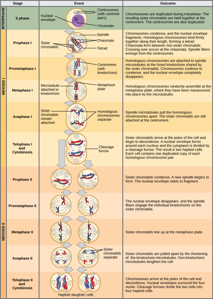
Stages of Meiosis
- Meiosis consists of two consecutive divisions: Meiosis I and Meiosis II. Each division has distinct phases analogous to those in mitosis but with unique events that contribute to genetic diversity.
Meiosis I: Reduction Division
- Objective: Reduce the chromosome number from diploid (2n) to haploid (n) by separating homologous chromosomes.
1. Prophase I
- Chromosome Condensation: Chromosomes condense, becoming visible under a microscope.
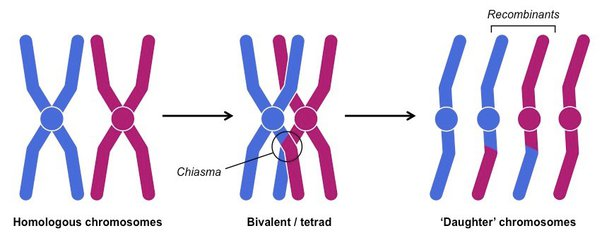
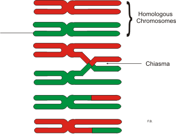
- Synapsis:
- Homologous Pairing: Each chromosome pairs with its homologous counterpart to form a bivalent or tetrad (a group of four chromatids).
- Crossing Over:
- Chiasmata Formation: Non-sister chromatids within the bivalent exchange genetic material at points called chiasmata, leading to genetic recombination.
- Nuclear Envelope Breakdown: The nuclear membrane disintegrates, allowing spindle fibers to interact with chromosomes.
- Spindle Fiber Attachment: Spindle fibers attach to the centromeres of each homologous chromosome.
Diagram: Illustration of a bivalent during Prophase I showing chiasmata.
Prometaphase I: The movement of the chromosomes to the metaplate/equator/middle of the cell is referred to as prometaphase.
2. Metaphase I
- Alignment at Equator: Bivalents align along the metaphase plate (cell equator).
- Independent Assortment: Homologous pairs orient randomly, contributing to genetic variation by mixing maternal and paternal chromosomes.
Diagram: Metaphase I showing randomly oriented bivalents.
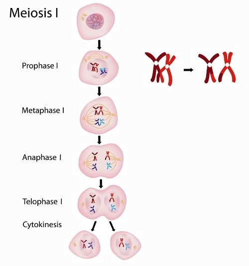
3. Anaphase I
- Separation of Homologs: Spindle fibers shorten, pulling homologous chromosomes to opposite poles of the cell.
- Centromere Integrity: Unlike mitosis, centromeres do not split; each homolog remains intact.
Diagram: Anaphase I illustrating homologous chromosomes moving to opposite poles.
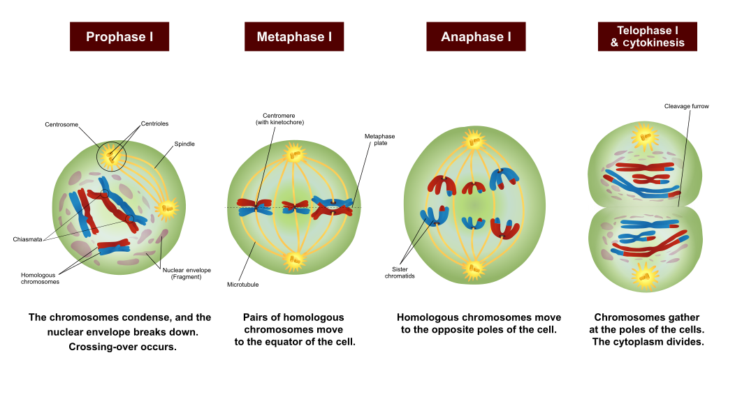
4. Telophase I and Cytokinesis
- Chromosome Arrival: Homologous chromosomes reach the poles.
- Nuclear Envelope Reformation: May reform around each set of chromosomes (more prominent in animal cells).
- Cytokinesis: Division of the cytoplasm results in two haploid cells, each containing one set of chromosomes (still consisting of sister chromatids).
Diagram: Telophase I showing two haploid cells with duplicated chromosomes.
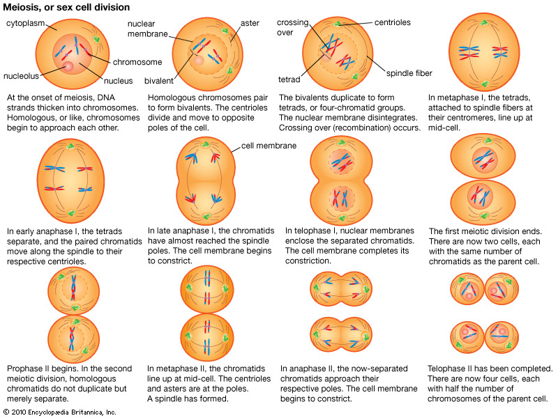
Meiosis II: Equational Division
- Objective: Separate sister chromatids, similar to mitosis, resulting in four haploid cells.
1. Prophase II
- Spindle Formation: New spindle fibers form in each haploid cell.
- Chromosome Preparation: Chromosomes, each with two sister chromatids, condense again if they had decondensed during Telophase I.
- Prometaphase II: This describes the movement of the chromosomes to the metaplate of the cell.
2. Metaphase II
- Alignment at Equator: Chromosomes line up individually along the metaphase plate in each haploid cell.
- Diagram Suggestion: Metaphase II showing chromosomes aligned singly.
3. Anaphase II
- Sister Chromatid Separation: Centromeres split, and spindle fibers pull sister chromatids (now individual chromosomes) to opposite poles.
Diagram: Anaphase II depicting chromatids moving to opposite poles.
4. Telophase II and Cytokinesis
- Chromosome Arrival: Chromosomes reach the poles and begin to decondense.
- Nuclear Envelope Reformation: Nuclear membranes form around each set of chromosomes.
- Cytokinesis: The cytoplasm divides, resulting in four genetically distinct haploid cells.
Diagram: Telophase II showing four haploid cells with single chromosomes.
Key Terms
- Meiosis: A type of cell division that reduces the chromosome number by half, producing four genetically distinct haploid cells.
- Haploid (n): A cell containing a single set of chromosomes.
- Diploid (2n): A cell containing two sets of chromosomes, one from each parent.
- Homologous Chromosomes: Pairs of chromosomes that have the same structure and gene sequence but may carry different alleles.
- Bivalent/Tetrad: A pair of homologous chromosomes aligned together during Prophase I.
- Chiasma (plural: Chiasmata): The point where two homologous non-sister chromatids exchange genetic material during crossing over.
- Crossing Over: The exchange of genetic material between non-sister chromatids, increasing genetic diversity.
- Independent Assortment: The random orientation of homologous chromosome pairs during Metaphase I, leading to varied combinations of maternal and paternal chromosomes in gametes.
- Reduction Division: The first division in meiosis that halves the chromosome number from diploid to haploid.
- Sister Chromatids: Identical copies of a single chromosome connected by a centromere.
Meiosis in Polyploid Cells
- Definition: Polyploid cells contain more than two sets of chromosomes (e.g., triploid 3n, tetraploid 4n).
- Challenges:
- Homologous Pairing Issues: In polyploid cells, homologous chromosomes may not align properly, making accurate segregation difficult.
- Outcome:
- Disrupted Reduction: The presence of multiple homologous pairs can interfere with the reduction division, potentially leading to unequal distribution of chromosomes and genetic instability.
- Implications: Meiosis is less likely to proceed normally in polyploid cells, which can affect fertility and viability.
Meiosis vs. Mitosis in Drosophila (Fruit Fly)
Assumption: Diploid number (2n) is 8 chromosomes.
| Feature | Mitosis | Meiosis |
|---|---|---|
| Number of Divisions | 1 | 2 |
| Daughter Cells Produced | 2 identical diploid cells | 4 non-identical haploid cells |
| Chromosomes per Nucleus | 8 (diploid) | 4 (haploid) |
| Genetic Variation | No variation (clones) | High variation (due to crossing over and independent assortment) |
Discussion Questions
- Stages of Meiosis:
- a. Homologous chromosomes pair: Prophase I
- b. Crossing over occurs: Prophase I
- c. Homologous chromosomes separate: Anaphase I
- d. Centromeres split, chromatids separate: Anaphase II
- e. Haploid nuclei form: Telophase I
- Meiosis in Polyploid Cells:
- Question: Why is meiosis unlikely to proceed normally in polyploid cells (e.g., 3n or 4n)?
- Answer: In polyploid cells, the presence of multiple homologous chromosome sets complicates the alignment and segregation processes. This improper pairing can disrupt the reduction division, leading to unequal chromosome distribution and genetic instability, making successful meiosis less likely.
Practice Questions
Question 1
Define meiosis and explain its primary purpose in sexually reproducing organisms.
Marking Scheme:
- Definition of Meiosis (2 marks):
- 1 mark: Correctly identifies meiosis as a type of cell division.
- 1 mark: Mentions that it reduces the chromosome number by half, producing haploid cells.
- Primary Purpose (3 marks):
- 1 mark: States that meiosis is essential for forming gametes (sperm and eggs) in animals or spores in plants.
- 1 mark: Explains that it ensures the maintenance of chromosome number across generations.
- 1 mark: Notes that meiosis introduces genetic variation through processes like crossing over and independent assortment.
Total: 5 marks
Answer Explanation: Meiosis is a type of cell division that reduces the chromosome number by half, producing haploid cells. Its primary purpose in sexually reproducing organisms is to form gametes (sperm and eggs) in animals or spores in plants, ensuring that when gametes fuse during fertilization, the diploid chromosome number is maintained. Additionally, meiosis introduces genetic variation through crossing over and independent assortment, which is crucial for evolution and adaptation.
Question 2
List and describe the four key differences between meiosis and mitosis.
Marking Scheme:
- Four Key Differences (4 marks):
- Number of Divisions:
- 1 mark: Mitosis involves one division cycle; meiosis involves two (Meiosis I & II).
- Purpose:
- 1 mark: Mitosis is for growth, repair, and asexual reproduction; meiosis is for producing gametes for sexual reproduction.
- Chromosome Number:
- 1 mark: Mitosis maintains the diploid number (2n); meiosis reduces the chromosome number by half (n).
- Genetic Variation:
- 1 mark: Mitosis produces genetically identical cells (no variation); meiosis increases genetic variation through crossing over and independent assortment.
- Number of Divisions:
Total: 4 marks
Answer Explanation:
- Number of Divisions: Mitosis consists of one division cycle, resulting in two identical diploid cells. Meiosis involves two consecutive division cycles (Meiosis I and II), producing four non-identical haploid cells.
- Purpose: Mitosis is primarily for growth, repair, and asexual reproduction, whereas meiosis is specialized for producing gametes necessary for sexual reproduction.
- Chromosome Number: Mitosis maintains the diploid chromosome number (2n) in the daughter cells. In contrast, meiosis reduces the chromosome number by half (n), ensuring gametes have a single set of chromosomes.
- Genetic Variation: Mitosis produces genetically identical cells with no variation. Meiosis introduces genetic diversity through mechanisms like crossing over during Prophase I and independent assortment during Metaphase I.
Question 3
During which phase of meiosis does crossing over occur, and why is it important for genetic variation?
Marking Scheme:
- Phase Identification (1 mark):
- Correct Phase: Prophase I of Meiosis I.
- Explanation of Importance (3 marks):
- 1 mark: Defines crossing over as the exchange of genetic material between non-sister chromatids.
- 1 mark: Explains that this exchange results in new combinations of alleles on each chromosome.
- 1 mark: States that crossing over increases genetic variation among offspring, which is beneficial for evolution and adaptation.
Total: 4 marks
Answer Explanation: Crossing over occurs during Prophase I of Meiosis I. It involves the exchange of genetic material between non-sister chromatids of homologous chromosomes, leading to new allele combinations. This process is crucial for generating genetic diversity, ensuring that offspring have unique genetic makeups, which enhances the adaptability and survival of populations.
Question 4
Compare the outcomes of mitosis and meiosis in terms of the number and genetic similarity of the resulting cells.
Marking Scheme:
- Number of Cells Produced (2 marks):
- Mitosis: Two identical diploid cells.
- Meiosis: Four non-identical haploid cells.
- Genetic Similarity (2 marks):
- Mitosis: Resulting cells are genetically identical to the parent cell (clones).
- Meiosis: Resulting cells are genetically diverse due to crossing over and independent assortment.
Total: 4 marks
Answer Explanation: Mitosis results in two identical diploid cells that are clones of the original parent cell, maintaining genetic similarity. In contrast, meiosis produces four non-identical haploid cells, each with unique genetic combinations due to processes like crossing over and independent assortment, thereby increasing genetic diversity.
Question 5
Explain the role of independent assortment during Metaphase I of meiosis and its contribution to genetic variation.
Marking Scheme:
- Definition of Independent Assortment (1 mark):
- States that it is the random orientation of homologous chromosome pairs at the metaphase plate.
- Mechanism Explanation (2 marks):
- 1 mark: Describes how homologous pairs align randomly, meaning maternal and paternal chromosomes are assorted into daughter cells independently of each other.
- 1 mark: Notes that this randomness leads to different combinations of chromosomes being distributed to each gamete.
- Contribution to Genetic Variation (1 mark):
- Highlights that independent assortment increases genetic variation by producing gametes with diverse genetic makeups.
Total: 4 marks
Answer Explanation: Independent assortment occurs during Metaphase I when homologous chromosome pairs align randomly along the metaphase plate. This random orientation means that each daughter cell receives a random mix of maternal and paternal chromosomes. As a result, the combination of chromosomes in each gamete is unique, significantly contributing to genetic variation among offspring.
Question 6
Describe what happens during Anaphase I of meiosis and how it differs from Anaphase in mitosis.
Marking Scheme:
- Process During Anaphase I (2 marks):
- 1 mark: Homologous chromosomes are separated and pulled to opposite poles of the cell by spindle fibers.
- 1 mark: Centromeres remain intact; sister chromatids do not separate.
- Difference from Mitosis (2 marks):
- 1 mark: In mitosis, sister chromatids are separated during Anaphase.
- 1 mark: In meiosis I, homologous chromosomes are separated, not sister chromatids.
Total: 4 marks
Answer Explanation: During Anaphase I of meiosis, homologous chromosomes are pulled apart to opposite poles of the cell by spindle fibers. Unlike mitosis, the centromeres do not split, so sister chromatids remain together. In contrast, during Anaphase of mitosis, the centromeres divide, and sister chromatids are separated into individual chromosomes, ensuring each daughter cell receives an identical set of chromosomes.
Question 7
What are the final products of meiosis II, and how do they compare to the products of meiosis I?
Marking Scheme:
- Final Products of Meiosis II (2 marks):
- Four genetically distinct haploid cells.
- Each cell contains single chromosomes (sister chromatids have separated).
- Comparison to Meiosis I (2 marks):
- Meiosis I: Produces two haploid cells, each with duplicated chromosomes (sister chromatids remain attached).
- Meiosis II: Further divides these two cells to produce four haploid cells, each with single chromosomes.
Total: 4 marks
Answer Explanation: Meiosis II results in four genetically distinct haploid cells, each containing individual chromosomes as sister chromatids have been separated. In contrast, meiosis I produces two haploid cells, each with duplicated chromosomes where sister chromatids remain joined. Thus, meiosis I reduces the chromosome number by half, and meiosis II ensures each haploid cell has single chromosomes, increasing genetic diversity.
Question 8
Why is meiosis less likely to proceed normally in polyploid cells (e.g., triploid or tetraploid)?
Marking Scheme:
- Explanation of Polyploidy (1 mark):
- Defines polyploid cells as having more than two sets of chromosomes (e.g., triploid 3n, tetraploid 4n).
- Challenges in Meiosis (2 marks):
- 1 mark: Homologous chromosomes may not align properly during pairing.
- 1 mark: Difficulty in accurate segregation due to multiple homologous pairs, leading to unequal distribution of chromosomes.
- Implications (1 mark):
- Leads to genetic instability and affects fertility and viability.
Total: 4 marks
Answer Explanation: In polyploid cells, which contain more than two sets of chromosomes, meiosis faces significant challenges in accurately pairing and segregating homologous chromosomes. The presence of multiple homologous pairs can disrupt the alignment and separation processes, resulting in unequal distribution of chromosomes to daughter cells. This genetic instability often impairs successful meiosis, negatively affecting fertility and the viability of the resulting gametes.
Question 9
Match the following stages with their correct descriptions in meiosis I: Prophase I, Metaphase I, Anaphase I, Telophase I.
Descriptions: A. Homologous chromosomes align at the cell equator. B. Homologous chromosomes are pulled to opposite poles. C. Chromosomes condense, synapse, and crossing over occurs. D. Nuclear envelope reforms and cytoplasm divides, resulting in two haploid cells.
Marking Scheme:
- Correct Matching (4 marks):
- Prophase I → C (1 mark)
- Metaphase I → A (1 mark)
- Anaphase I → B (1 mark)
- Telophase I → D (1 mark)
Total: 4 marks
Answer Explanation:
- Prophase I: Chromosomes condense, homologous chromosomes pair up (synapse), and crossing over occurs.
- Metaphase I: Homologous chromosome pairs align randomly at the metaphase plate (cell equator).
- Anaphase I: Homologous chromosomes are separated and pulled to opposite poles of the cell.
- Telophase I: The nuclear envelope reforms around each set of chromosomes, and cytokinesis divides the cell into two haploid cells.
Question 10
Illustrate and label the key events of Meiosis II, ensuring to include Prophase II, Metaphase II, Anaphase II, and Telophase II.
Marking Scheme:
Note: Since this is a written response, focus on key points that should be included in the illustration and labels.
- Prophase II (1 mark):
- New spindle fibers form.
- Chromosomes condense if they had decondensed.
- Metaphase II (1 mark):
- Chromosomes align individually along the metaphase plate in each haploid cell.
- Anaphase II (1 mark):
- Centromeres split.
- Sister chromatids are pulled to opposite poles.
- Telophase II (1 mark):
- Chromosomes reach the poles and begin to decondense.
- Nuclear envelopes reform around each set of chromosomes.
- Cytokinesis divides each cell, resulting in four haploid cells.
Total: 4 marks
Answer Explanation: An accurate illustration should depict each stage with chromosomes and spindle fibers appropriately labeled.
- Prophase II: Shows spindle fibers forming and chromosomes condensing.
- Metaphase II: Chromosomes line up singly along the metaphase plate in each haploid cell.
- Anaphase II: Sister chromatids are separated and pulled to opposite poles.
- Telophase II: Chromosomes arrive at the poles, nuclear envelopes reform, and cytokinesis results in four distinct haploid cells.
Summary of Key Concepts Covered:
- Purpose and Function of Meiosis: Production of gametes/spores, reduction of chromosome number, introduction of genetic variation.
- Differences Between Meiosis and Mitosis: Number of divisions, purpose, chromosome number maintenance, genetic variation, and outcomes.
- Stages of Meiosis I and II: Key events and their significance in cell division and genetic diversity.
- Genetic Variation Mechanisms: Crossing over and independent assortment.
- Challenges in Polyploid Cells: Impact on meiosis and genetic stability.
- Comparative Outcomes: Understanding the genetic similarity and diversity resulting from different types of cell division.